Diabetic Eye Disease and
Diabetic Retinopathy
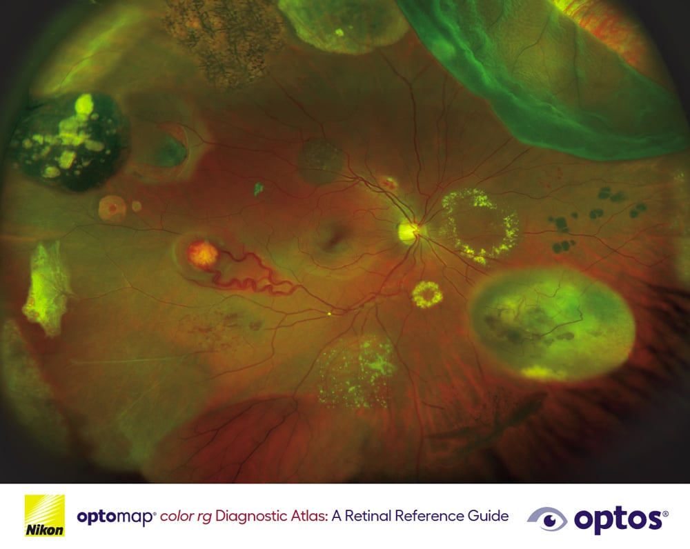
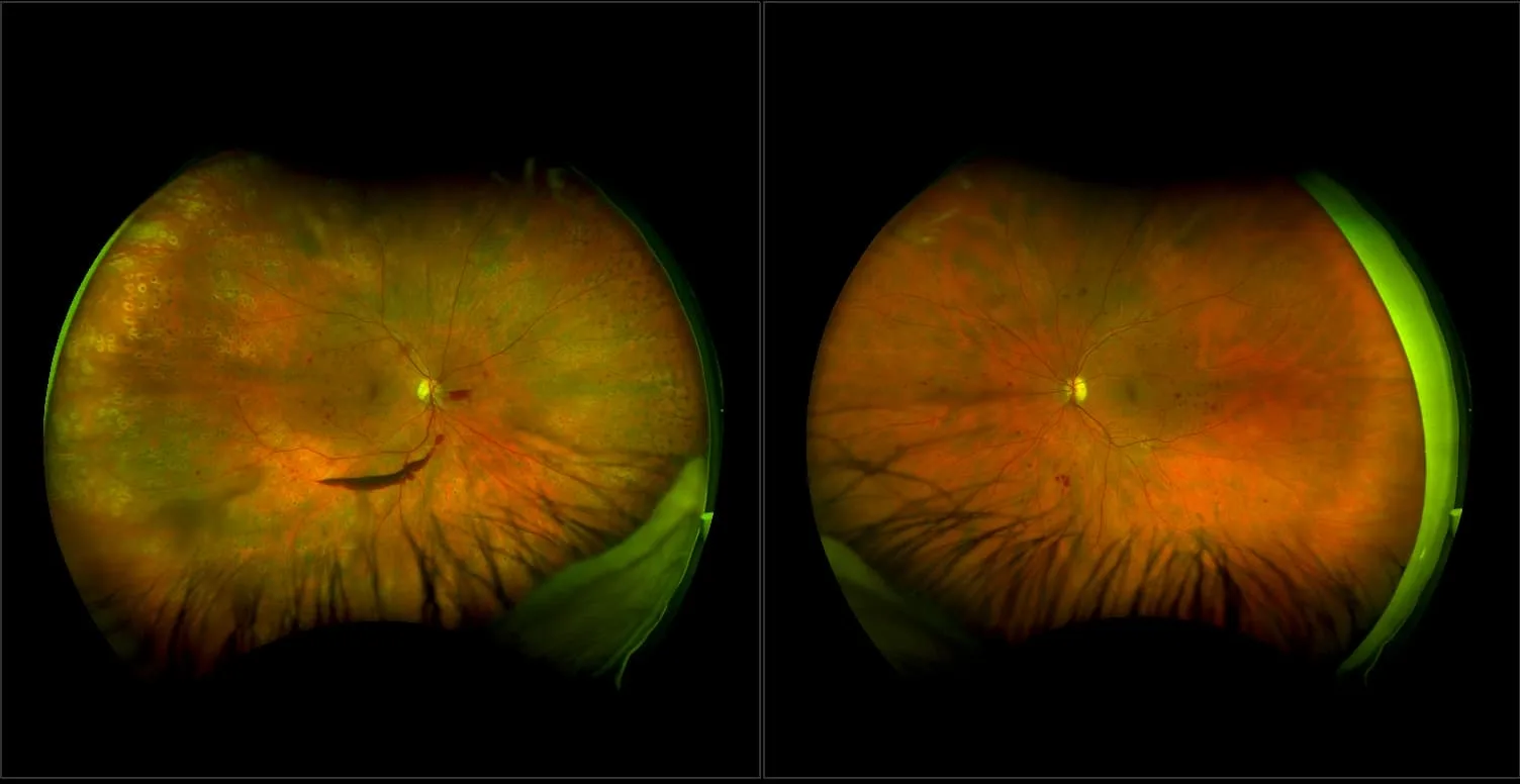
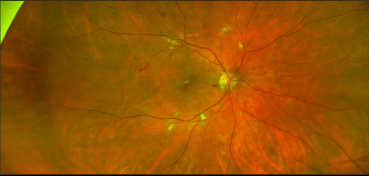
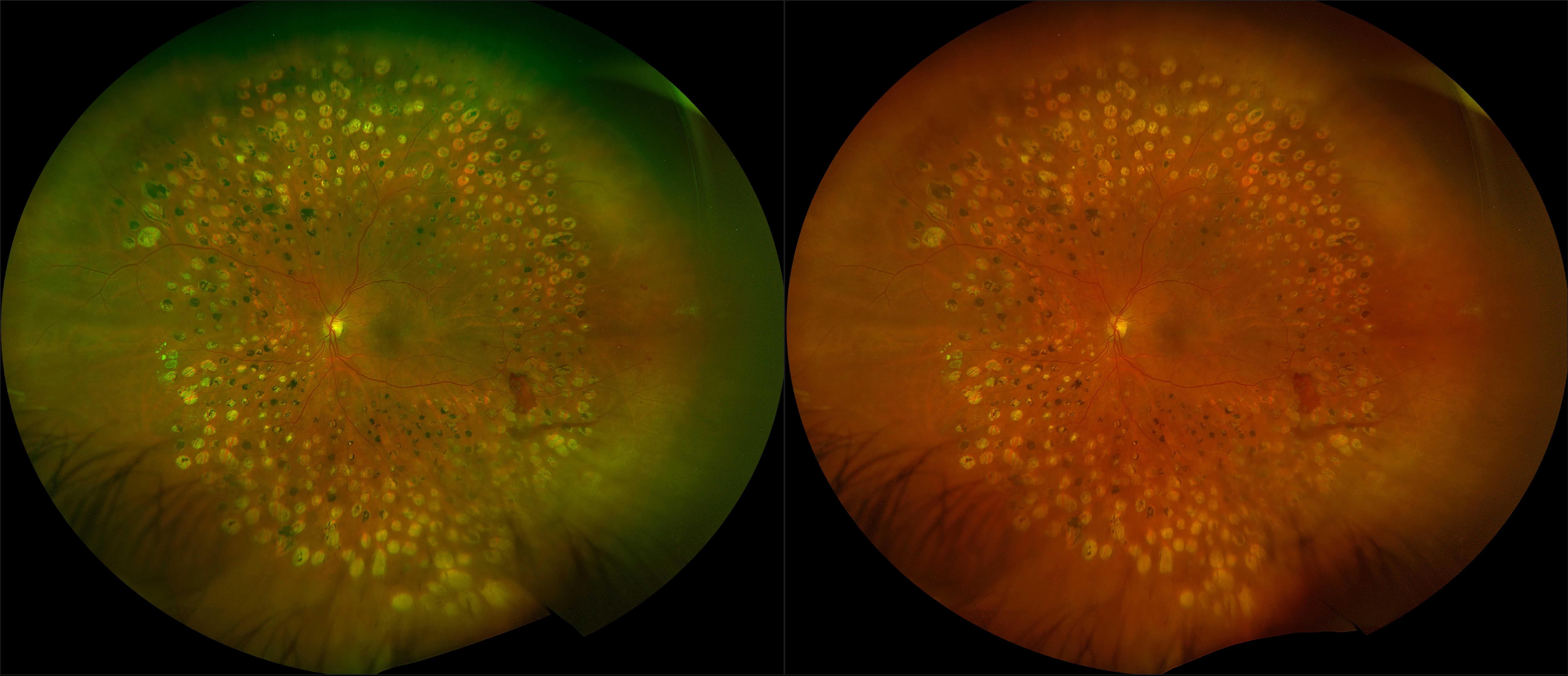
Diabetic eye disease is a complication of Diabetes Mellitus. Diabetic retinopathy is often detected as a complication, and it presents by affecting small blood vessels in the eye due to blockage or leakage. Over time, blood vessels can present as microaneurysms or hemorrhages or fluid (exudates). The number and severity of affected vessels determine the grade or retinopathy. There are 2 forms, nonproliferative (NPDR) and proliferative (PDR).
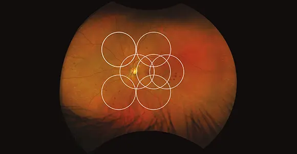
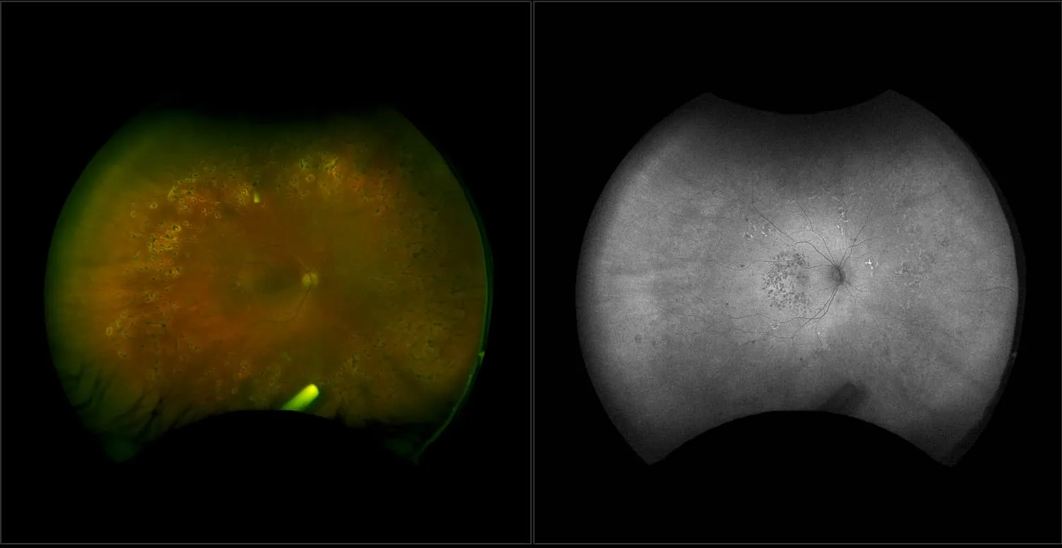
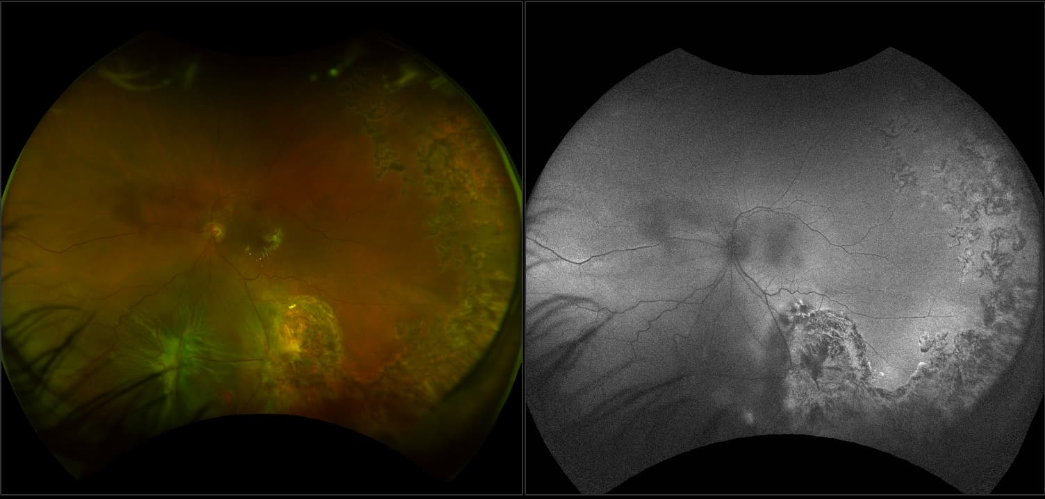
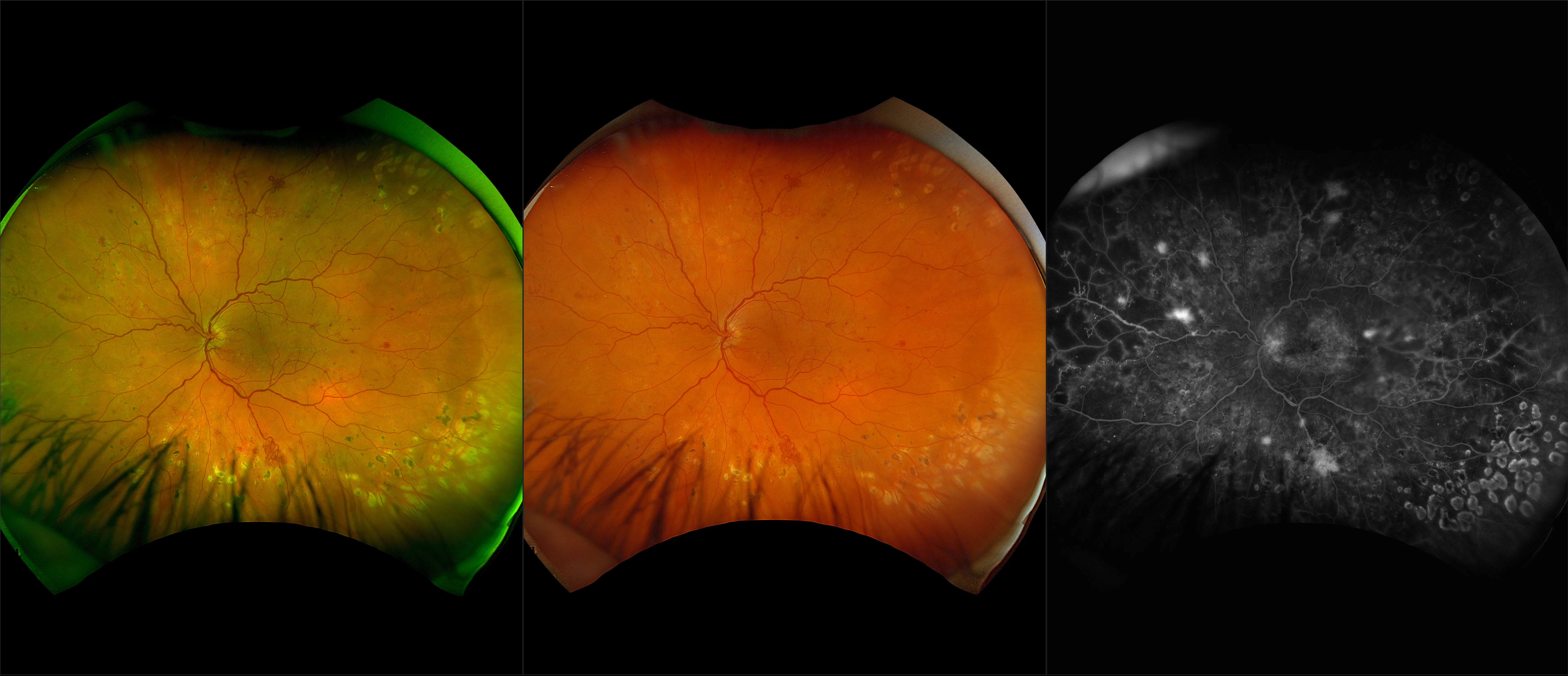
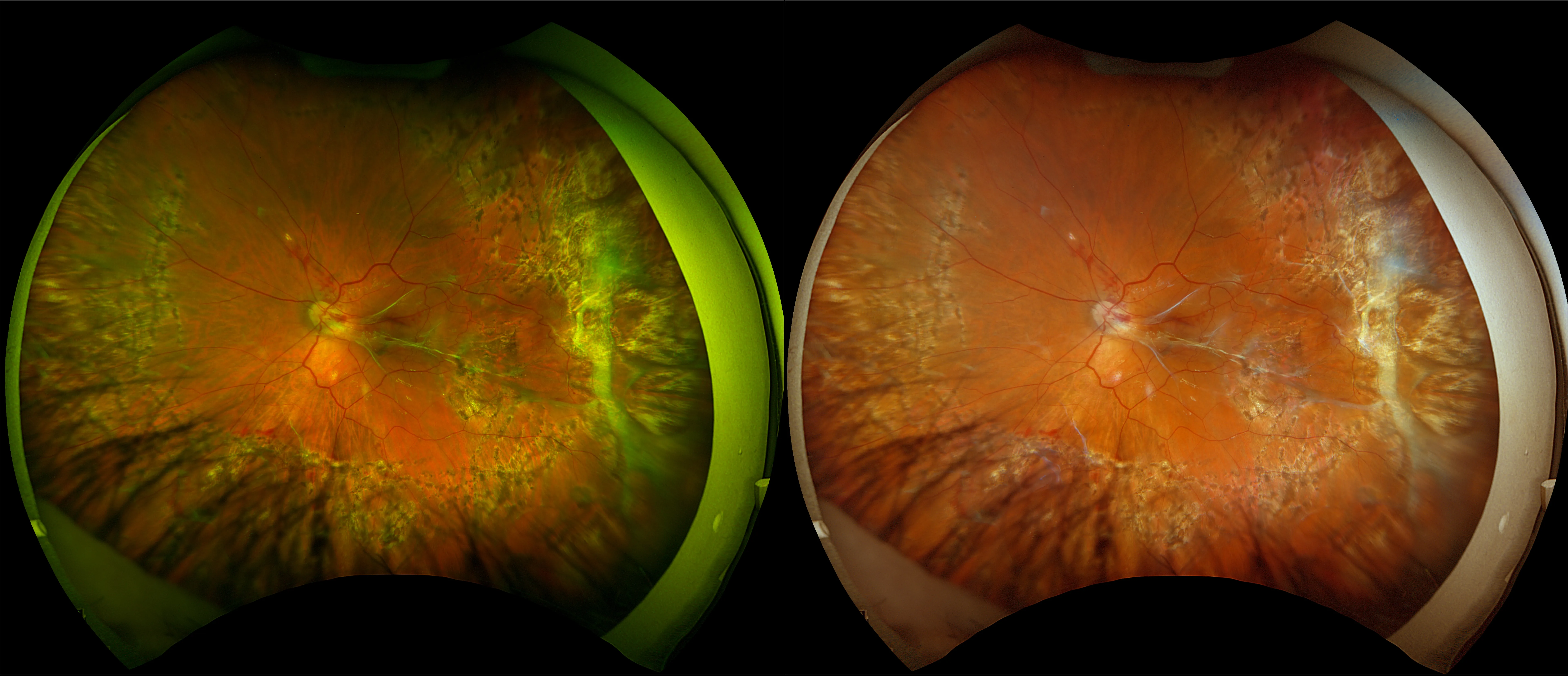
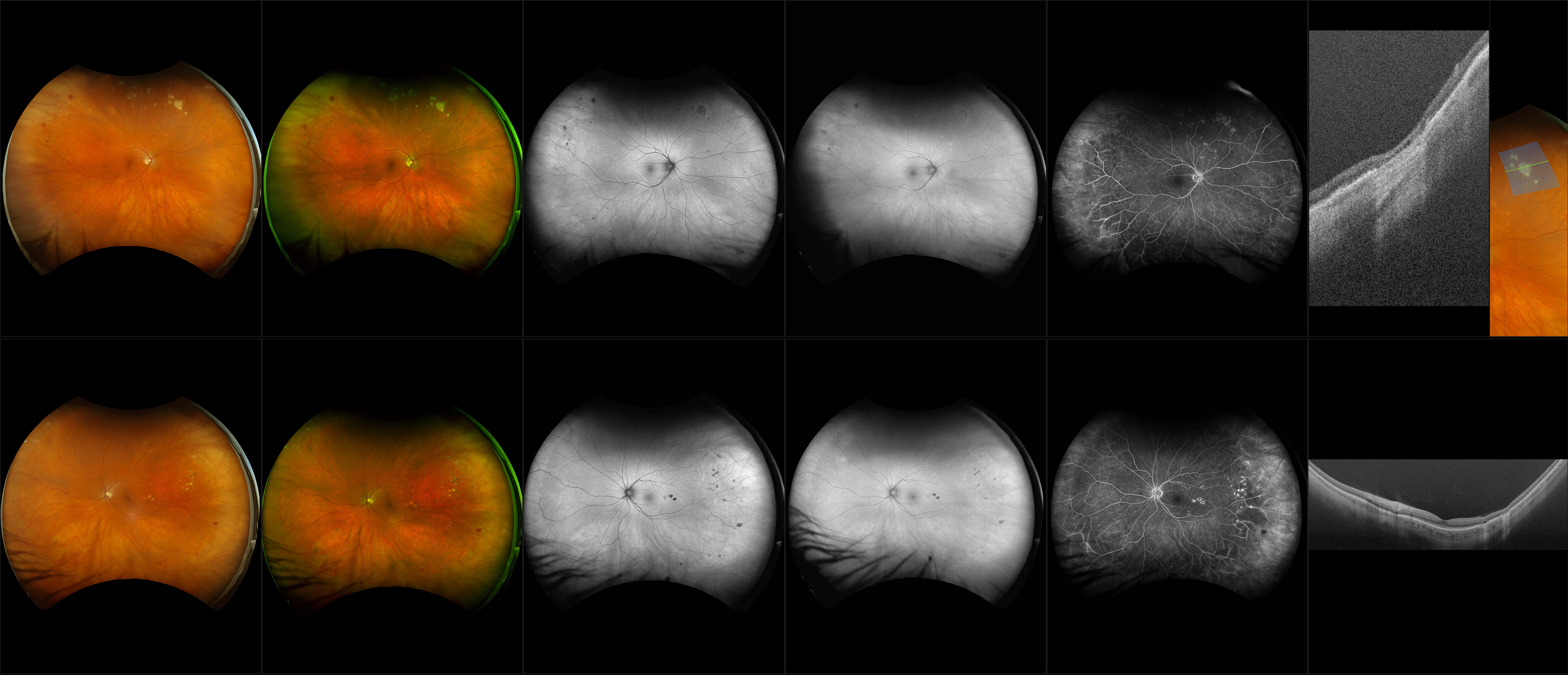
optomap imaging has been shown to improve the management of diabetes in patients. optomap images capture a 200° view (about 82%) of the retina versus the 75° view provided by 7SF images. Studies have shown that this wider view can uncover evidence of disease that is outside the narrow view of 7SF images. It can even change how doctors judge the severity of the disease. In addition, the latest study identified that 50% of the lesions were in the area outside of ETDRS and that in 13% of patients, these lesions suggested a more severe grade of retinopathy.
Evidence of disease at the periphery of the retina can also be a sign of future problems. Patients with peripheral DR lesions were more than four times more likely to see their DR get worse as compared to patients without lesions.
Diabetic retinopathy can impact people of all ages and in cases of diabetic eye diseases and further complications, early detection is key. optomap can help doctors to better monitor and facilitate decision making regarding treatment.
Clinical Summaries
optomap Equivalent to ETDRS
Studies confirm the equivalence of optomap to ETDRS Gold Standard for grading diabetic retinopathy.
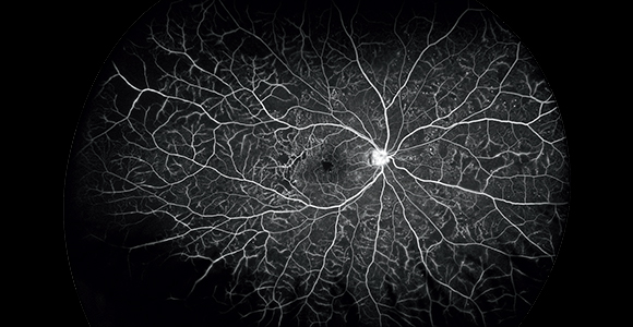
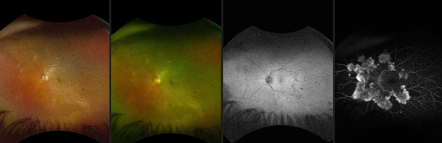
optomap fa Key Indicator to Predict the Progression of PDR
Research using optomap fa reveals that 50% of eyes with baseline PPL are at high risk for DR.
Referenced Papers
optomap Equivalent to ETDRS
Studies confirm the equivalence of optomap to ETDRS Gold Standard for grading diabetic retinopathy.
Peripheral Lesions Identified on Ultrawide Field Imaging Predict Increased Risk of Diabetic Retinopathy Progression over 4 Years
optomap imaging has demonstrated that diabetic lesions occur in the retinal periphery in up to 50% of eyes and these lesions might result in a more severe grade of retinopathy in 10% of eyes. Eyes with predominantly peripheral lesions (defined as outside of ETDRS 7 standard field) had a 4.7 fold increased risk of progression to proliferative diabetic retinopathy (PDR). Eyes with predominantly peripheral lesions had a 3.2 fold risk of 2 step progression in DR.
optomap for Telemedicine Programs
Ocular telemedicine programs that include optomap have a nearly double detection rate of DR.
optomap fa Key Indicator to Predict the Progression of PDR
Research using optomap fa reveals that 50% of eyes with baseline PPL are at high risk for DR.
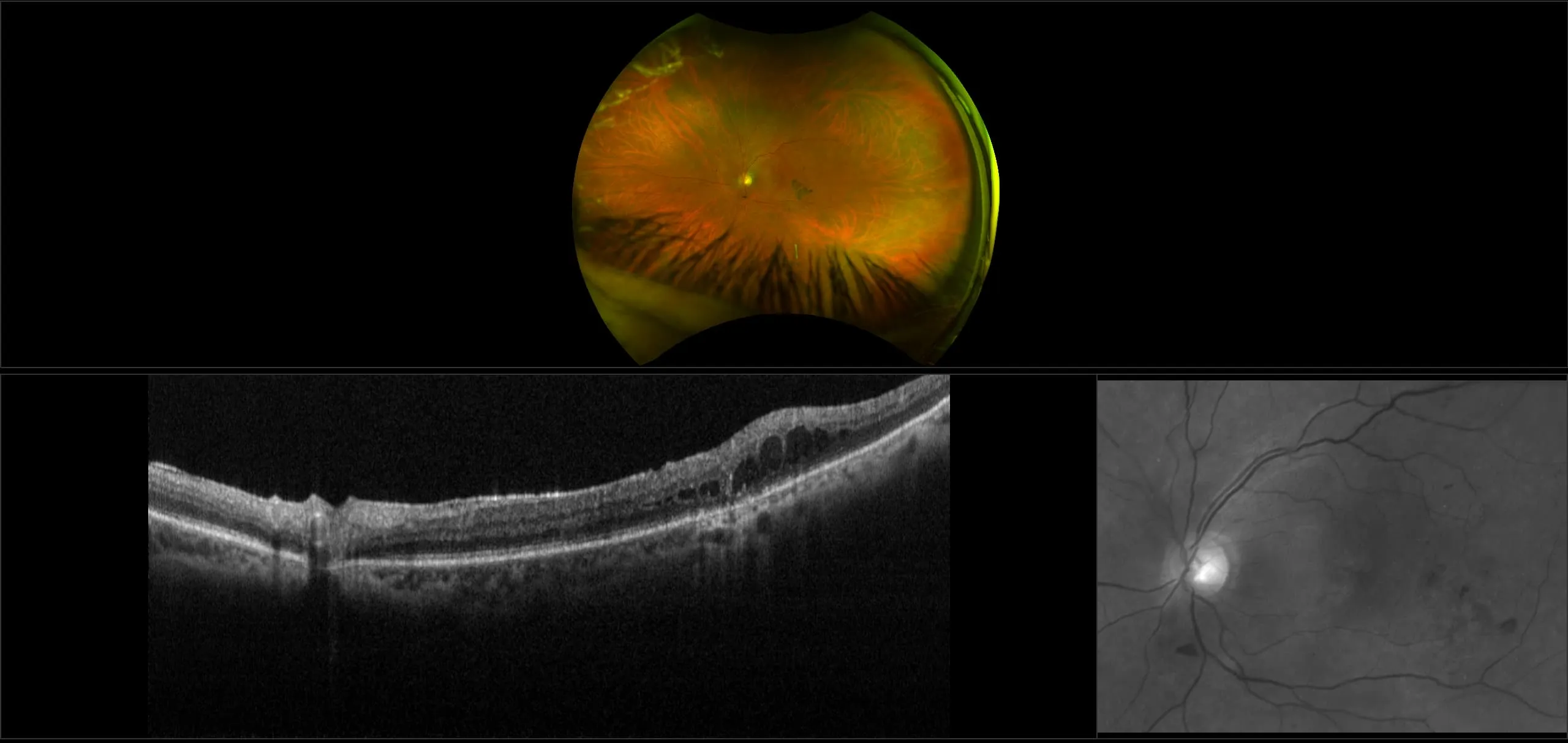
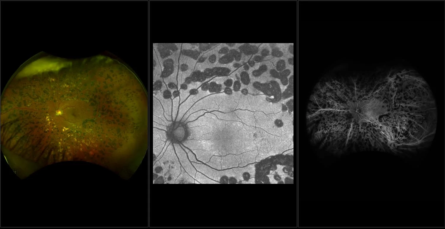
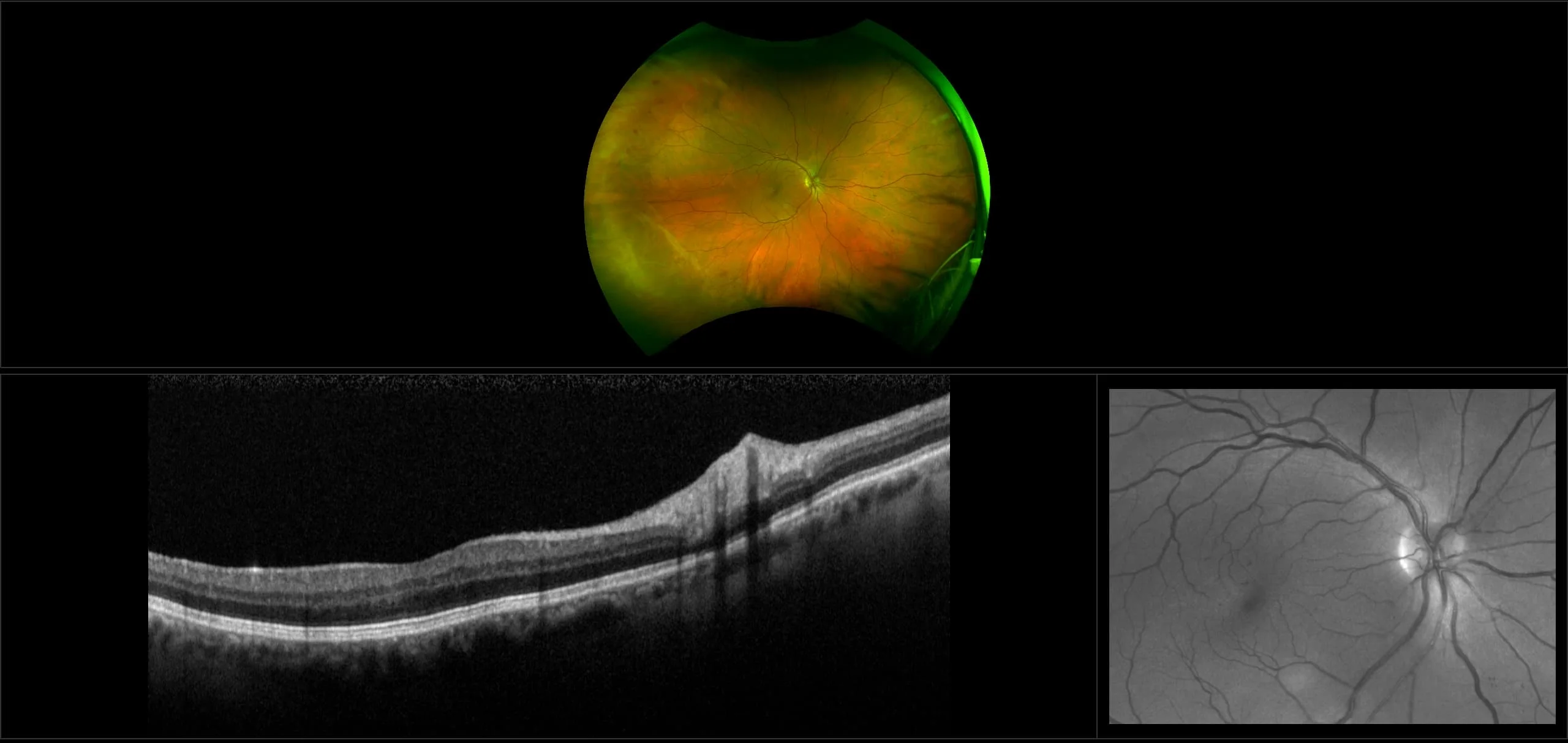
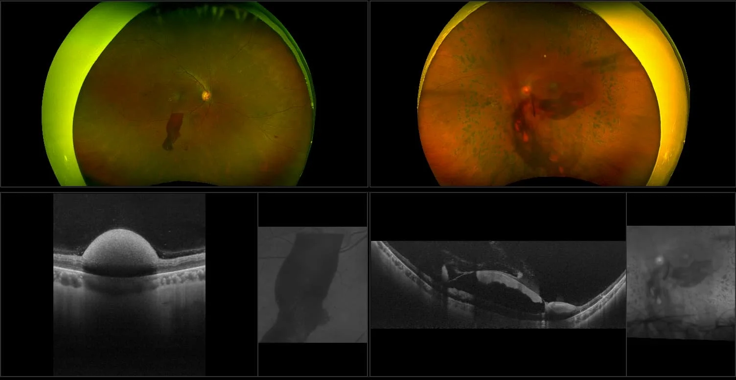
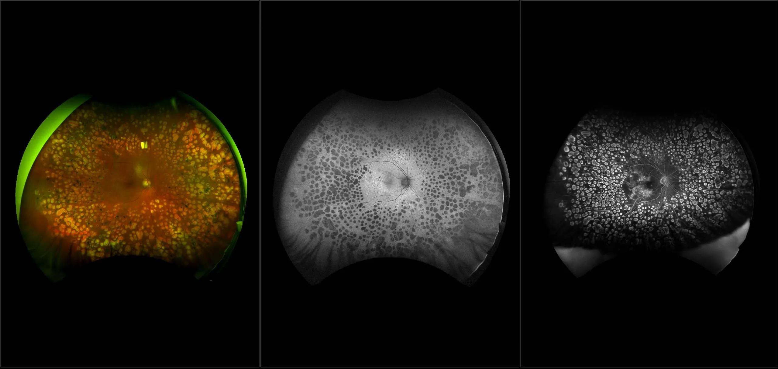
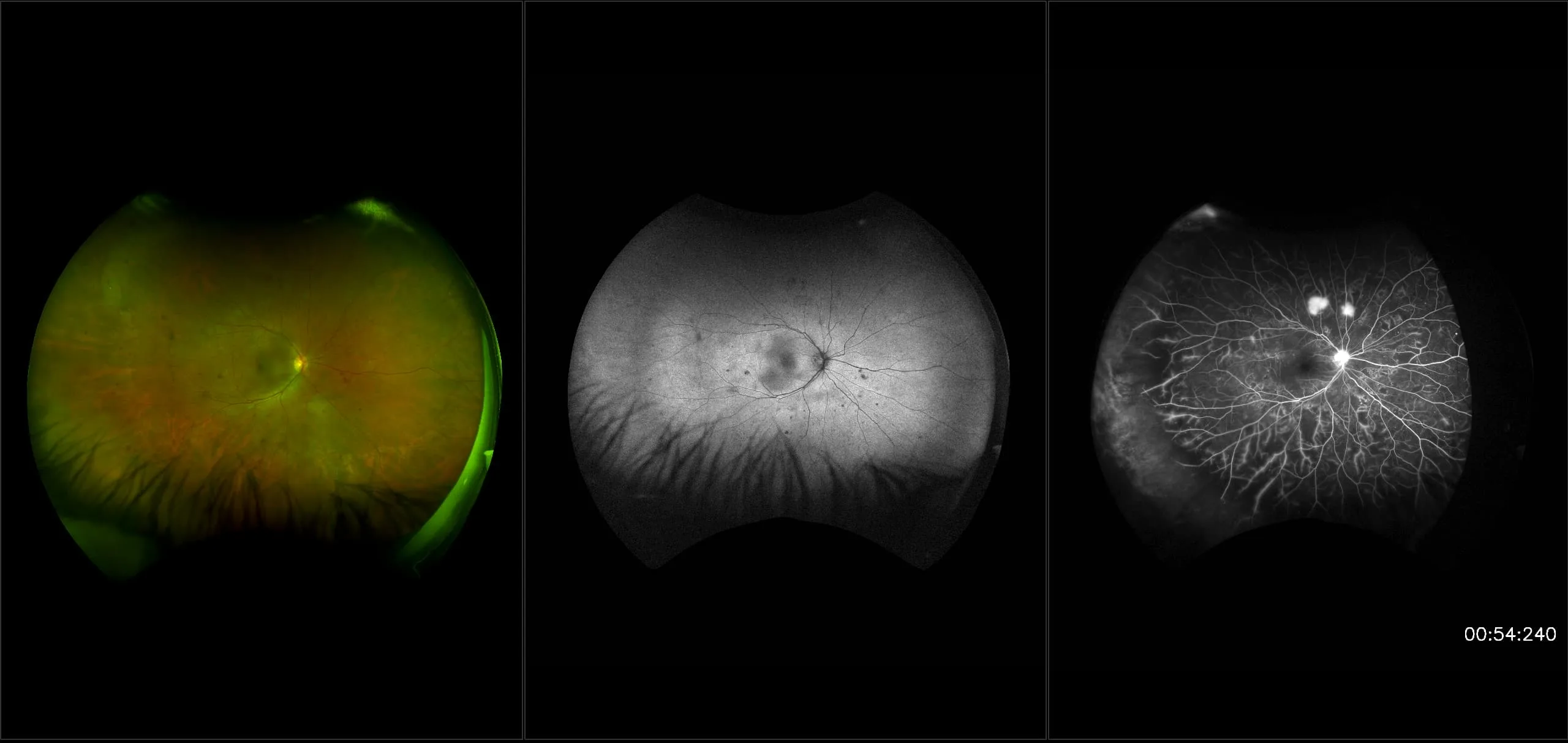
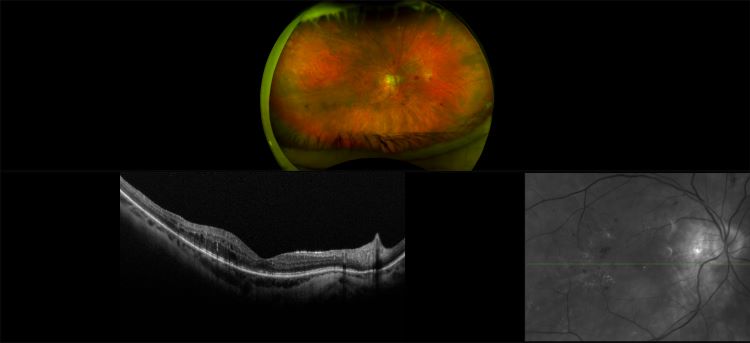
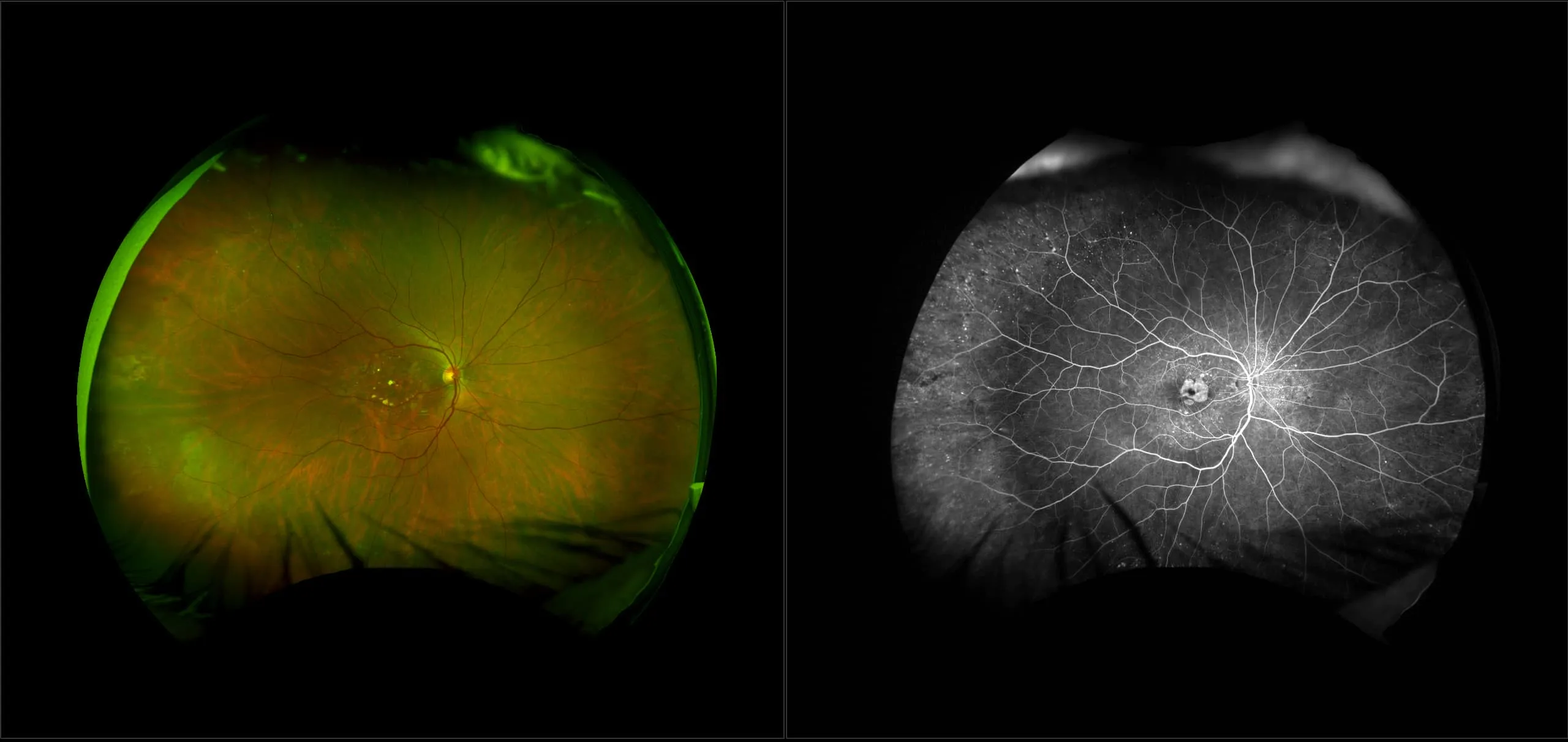
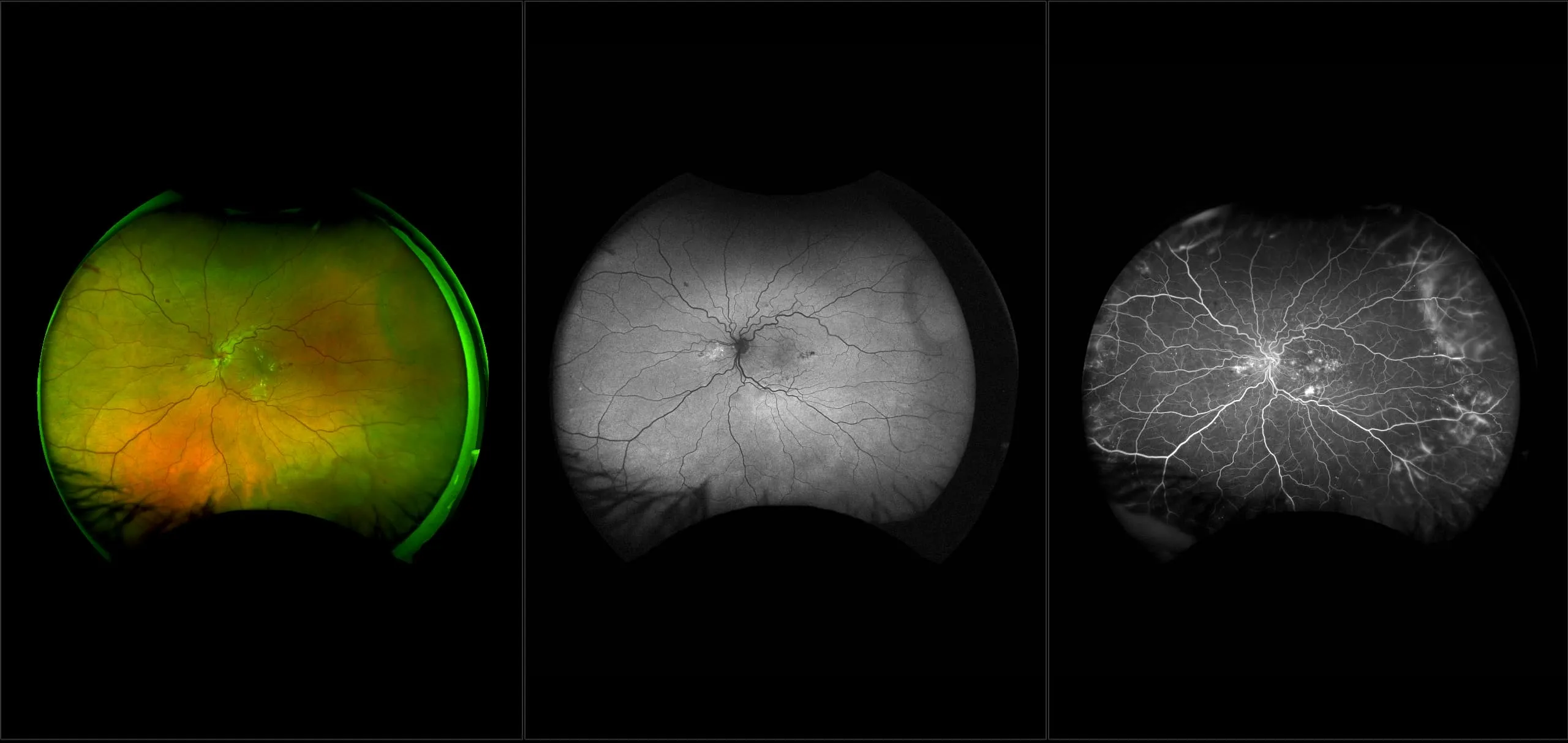
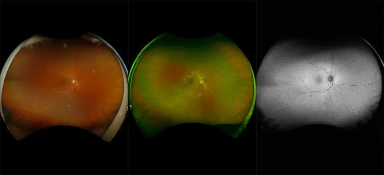
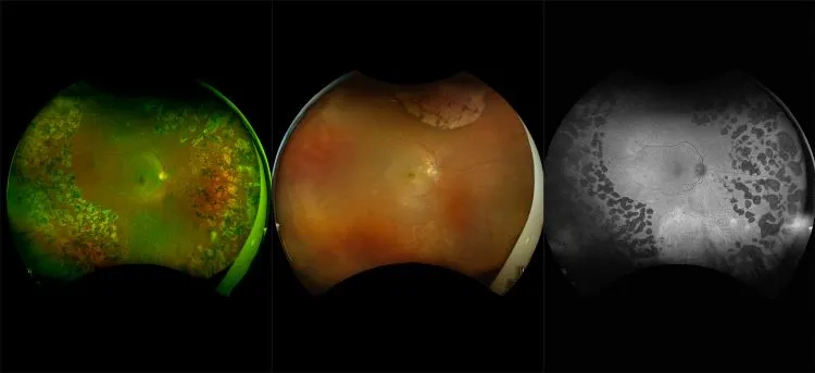
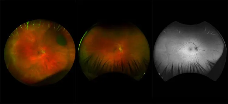
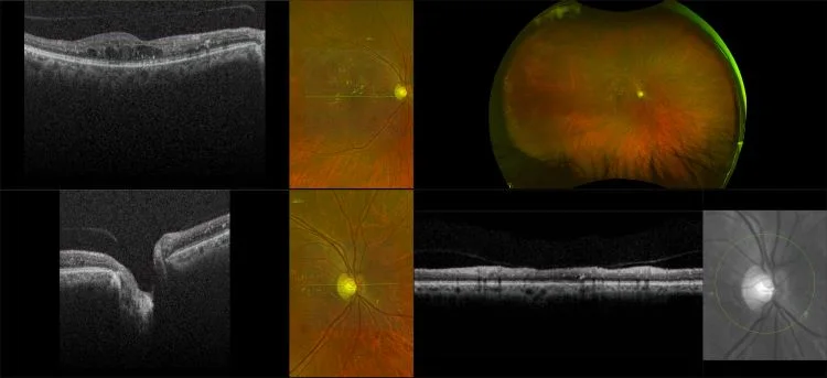
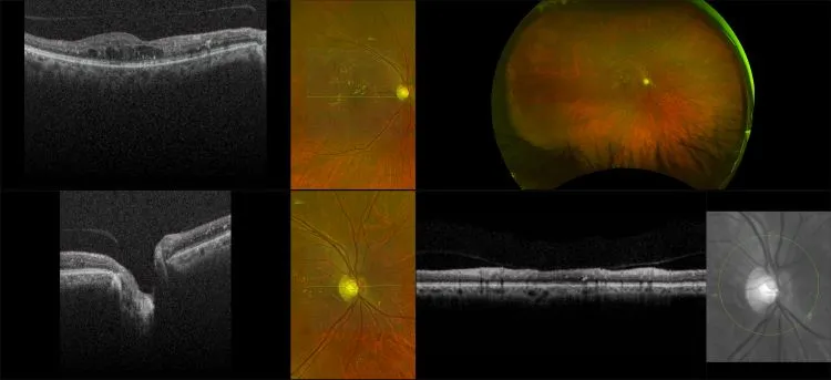
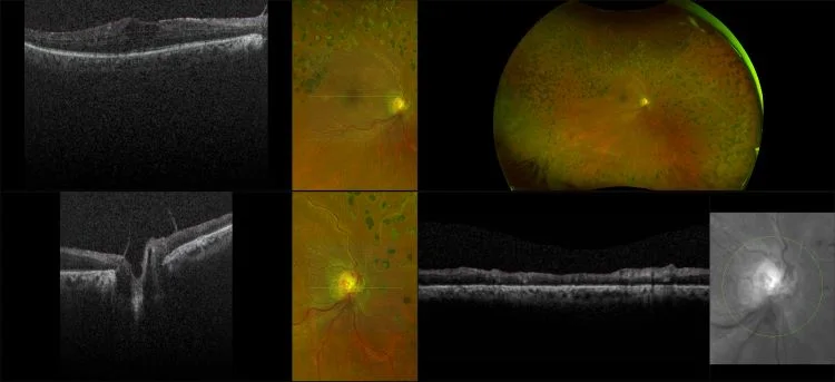
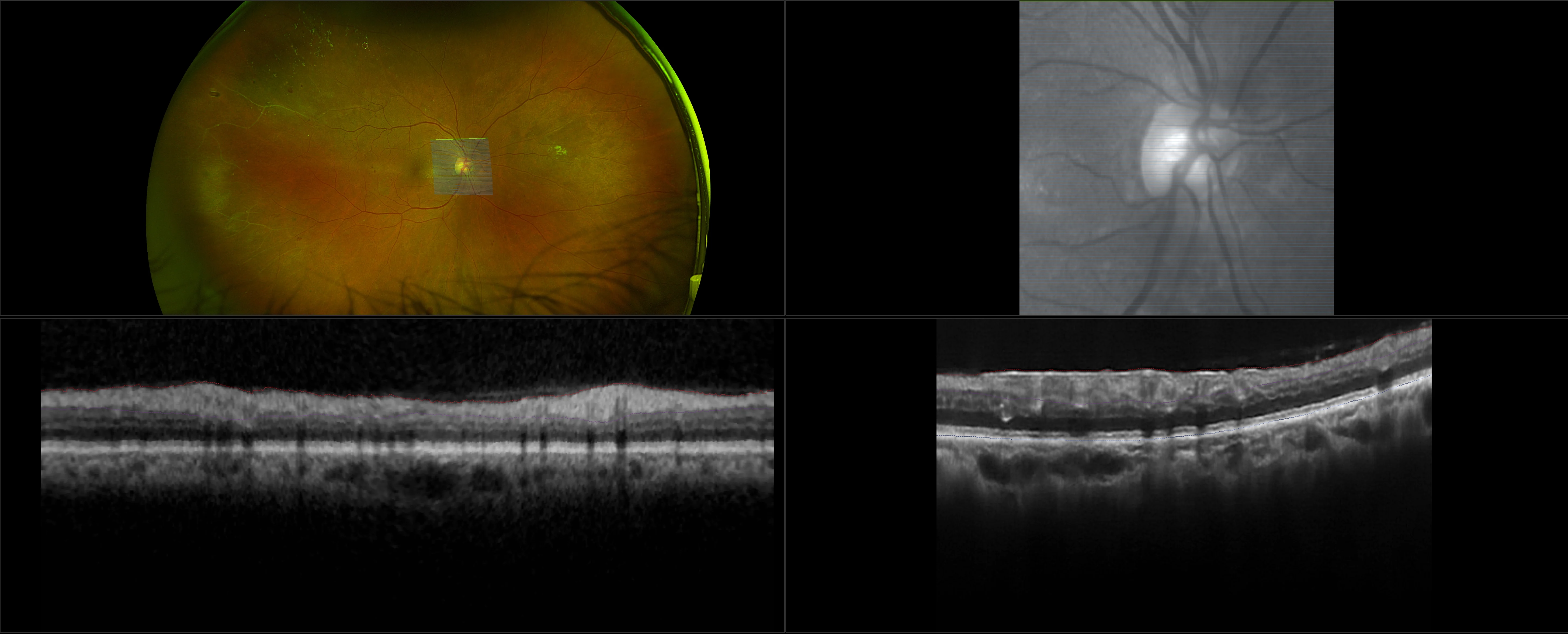
optomap Multimodal Diabetic Eye Disease Cases
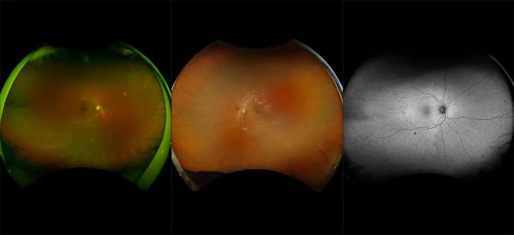
Optos offers multimodal imaging with all ultra-widefield devices. Having both ultra-widefield and four images captured in less than one second has been shown to enhance pathology detection and disease management as well as improve practice and clinic flow. Ultra-widefield multimodal imaging is important across all access points of patient care - screening, detection, diagnosis, and treatment.
Related Cases

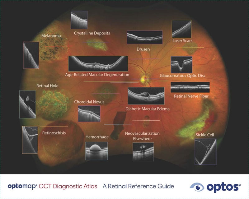
Other Diagnostic Tools