optomap® Recognizing Pathology
This material is designed as a searchable reference resource to support clinical decision-making. The information contained here should be used as general guidance when viewing optomap and OCT images from Optos devices. The differential diagnosis should be made under the direction of the responsible physician. These images were taken on the latest ultra-widefield optomap devices.
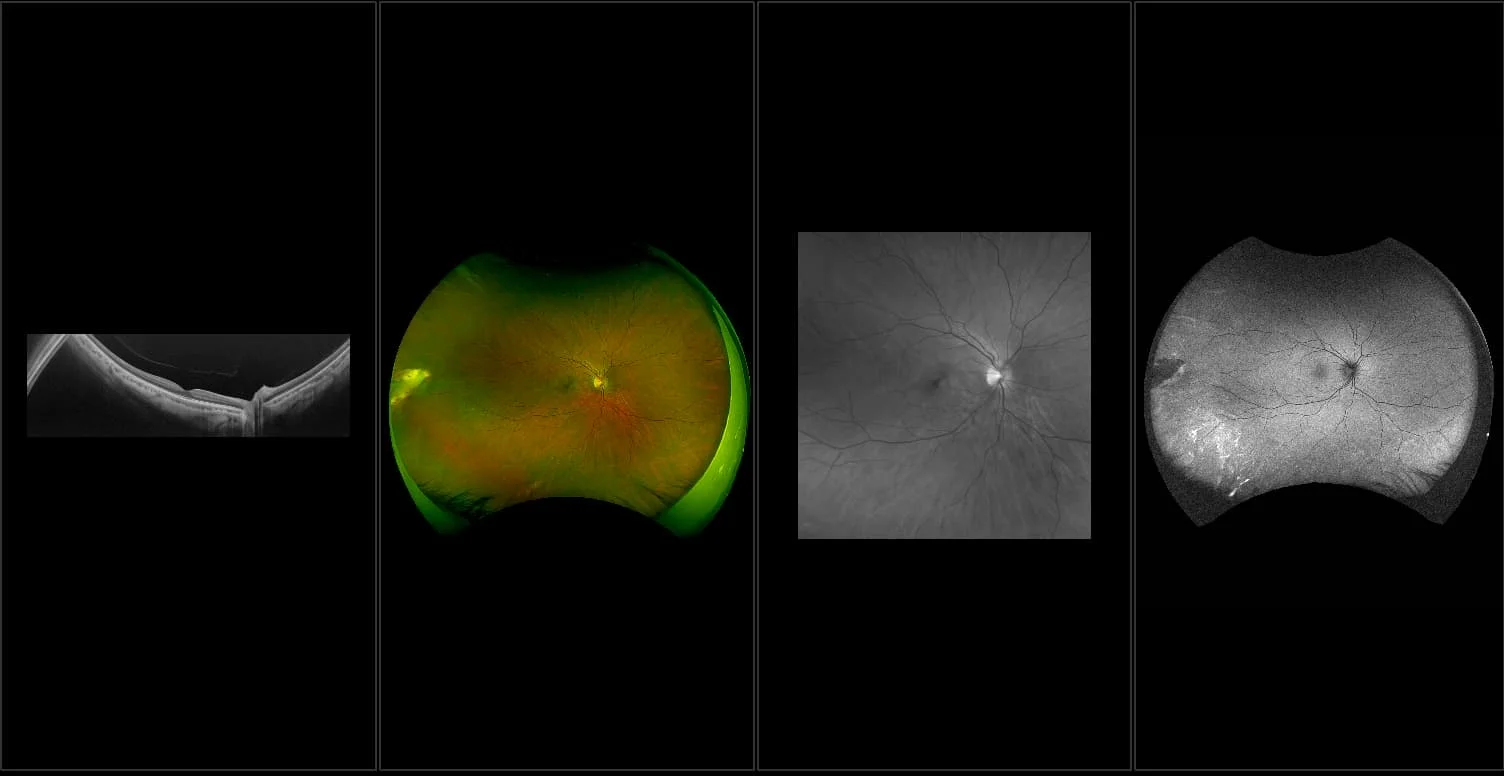
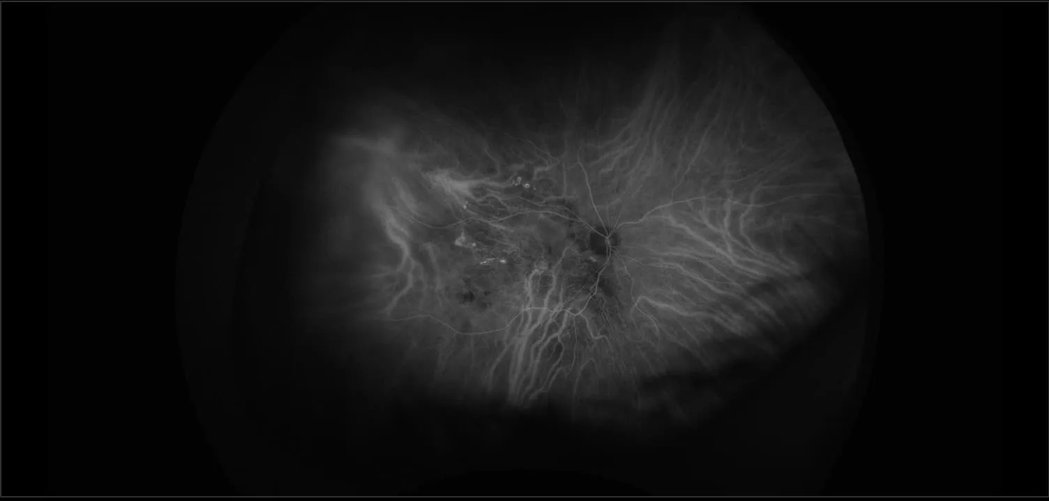
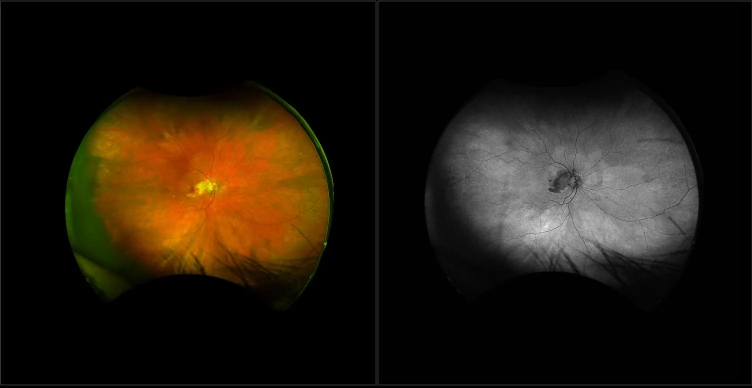
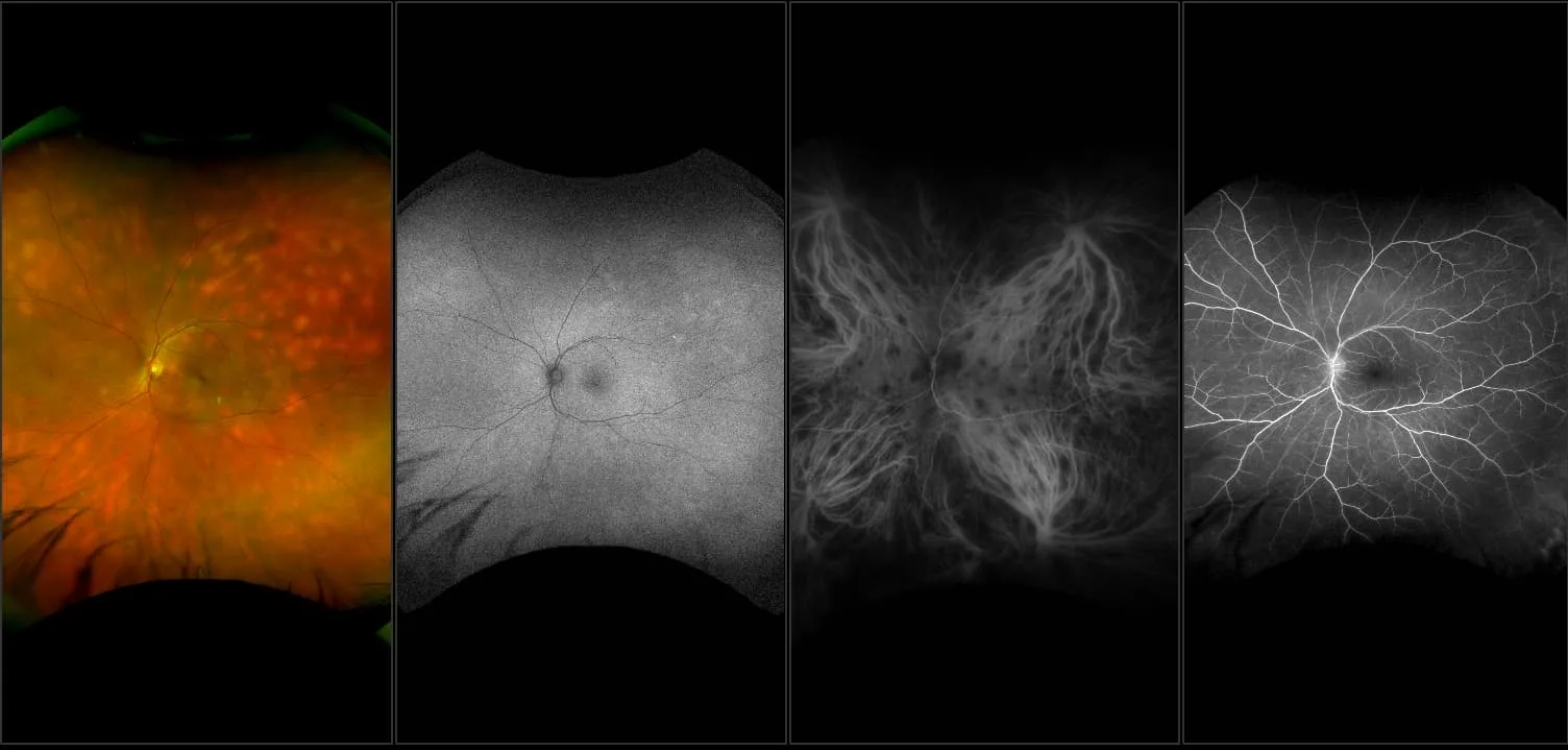
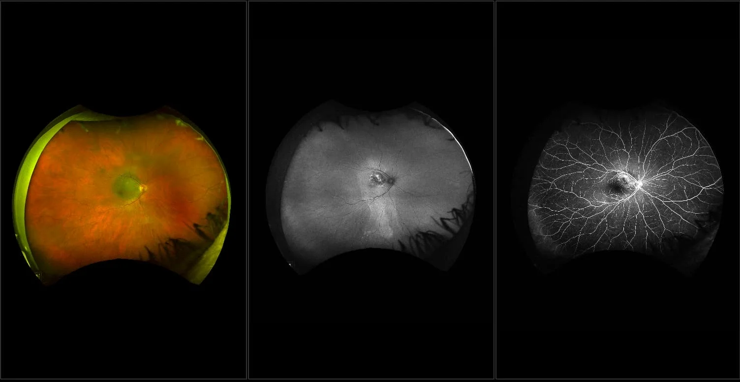
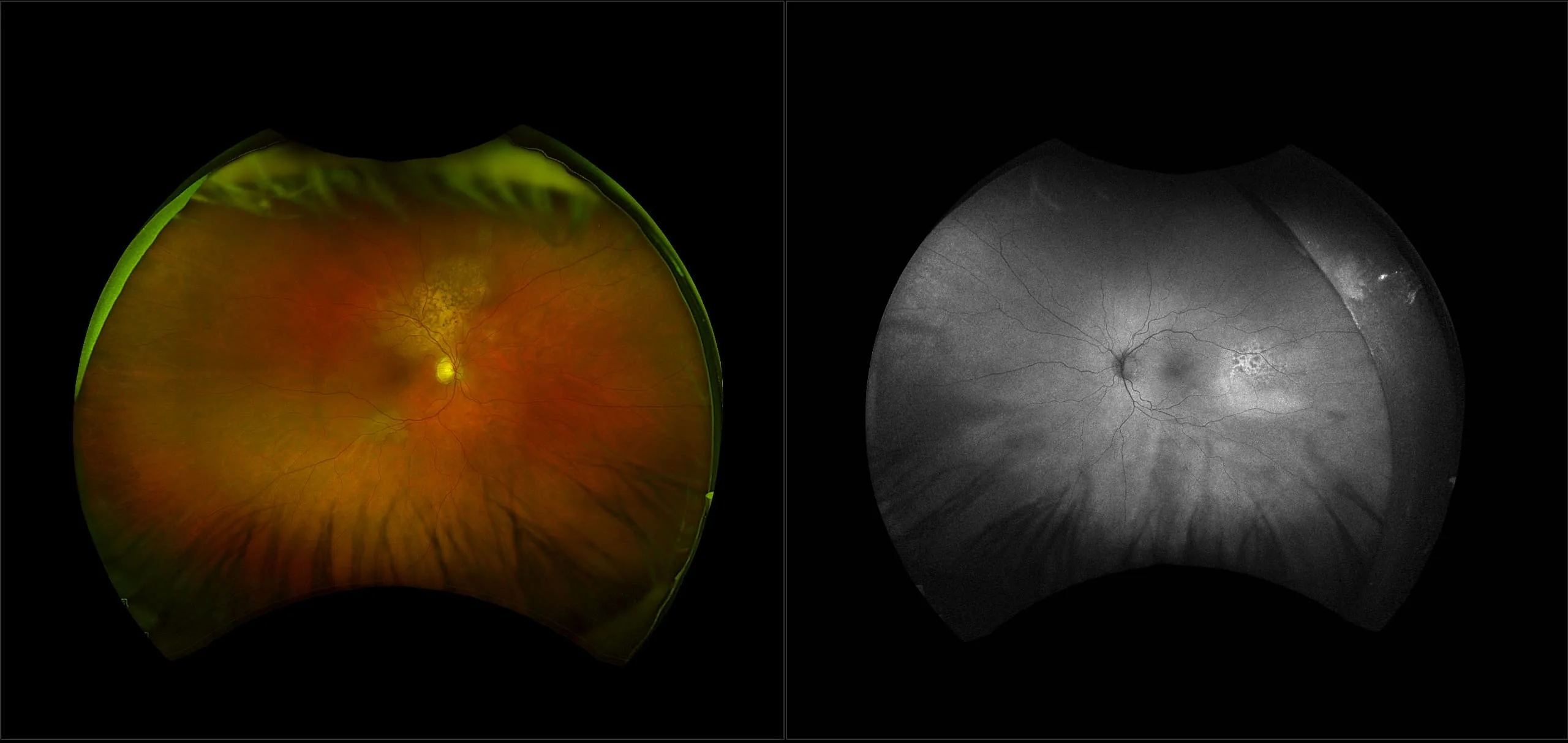
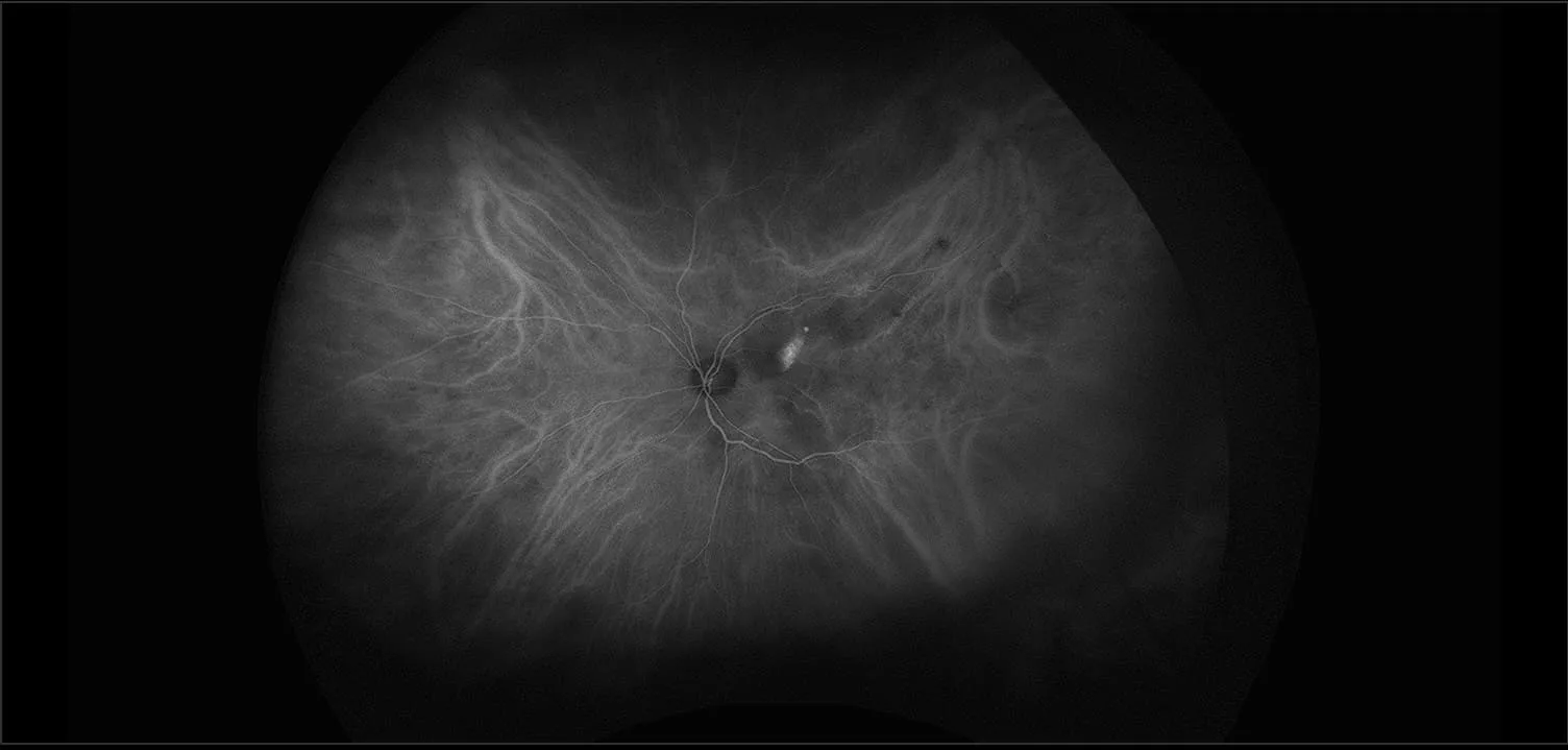
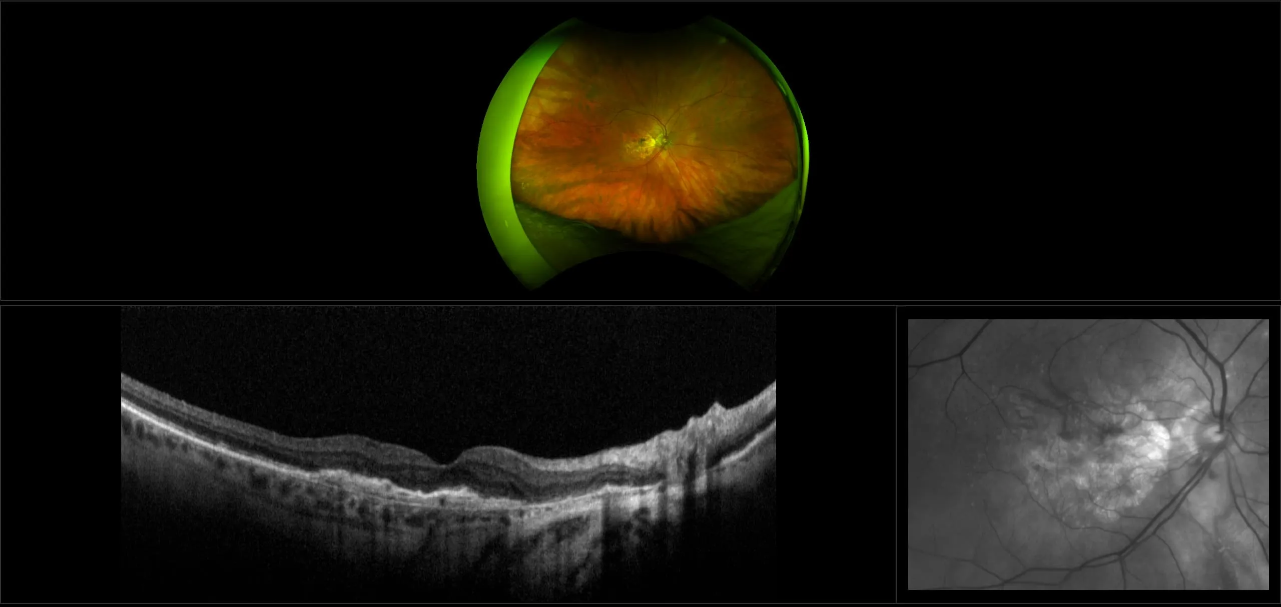
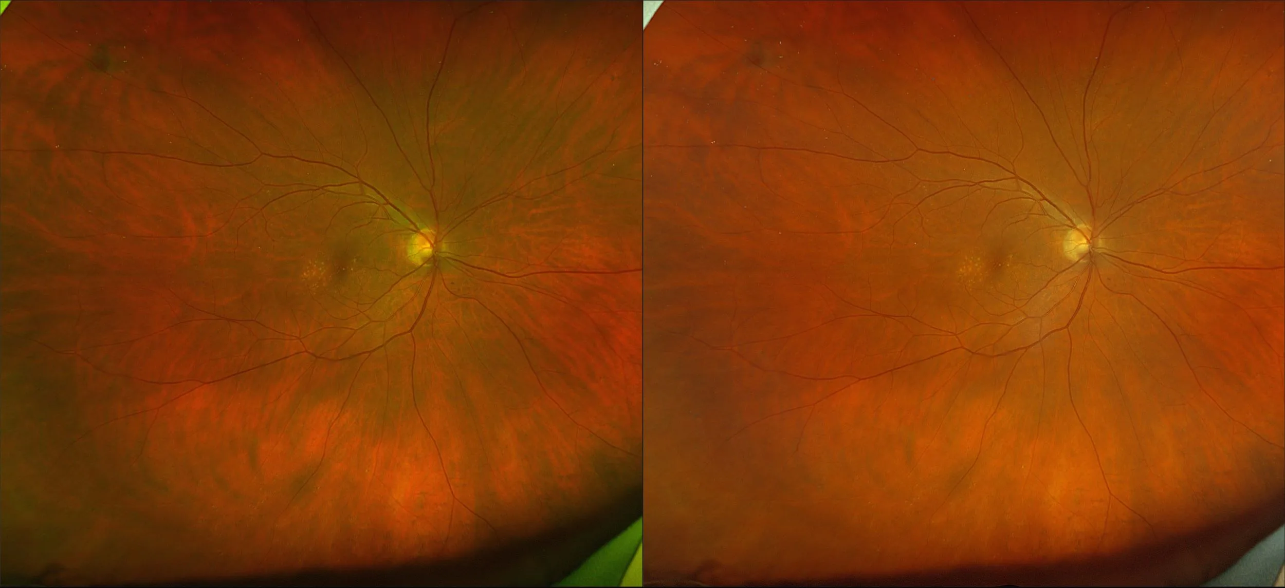
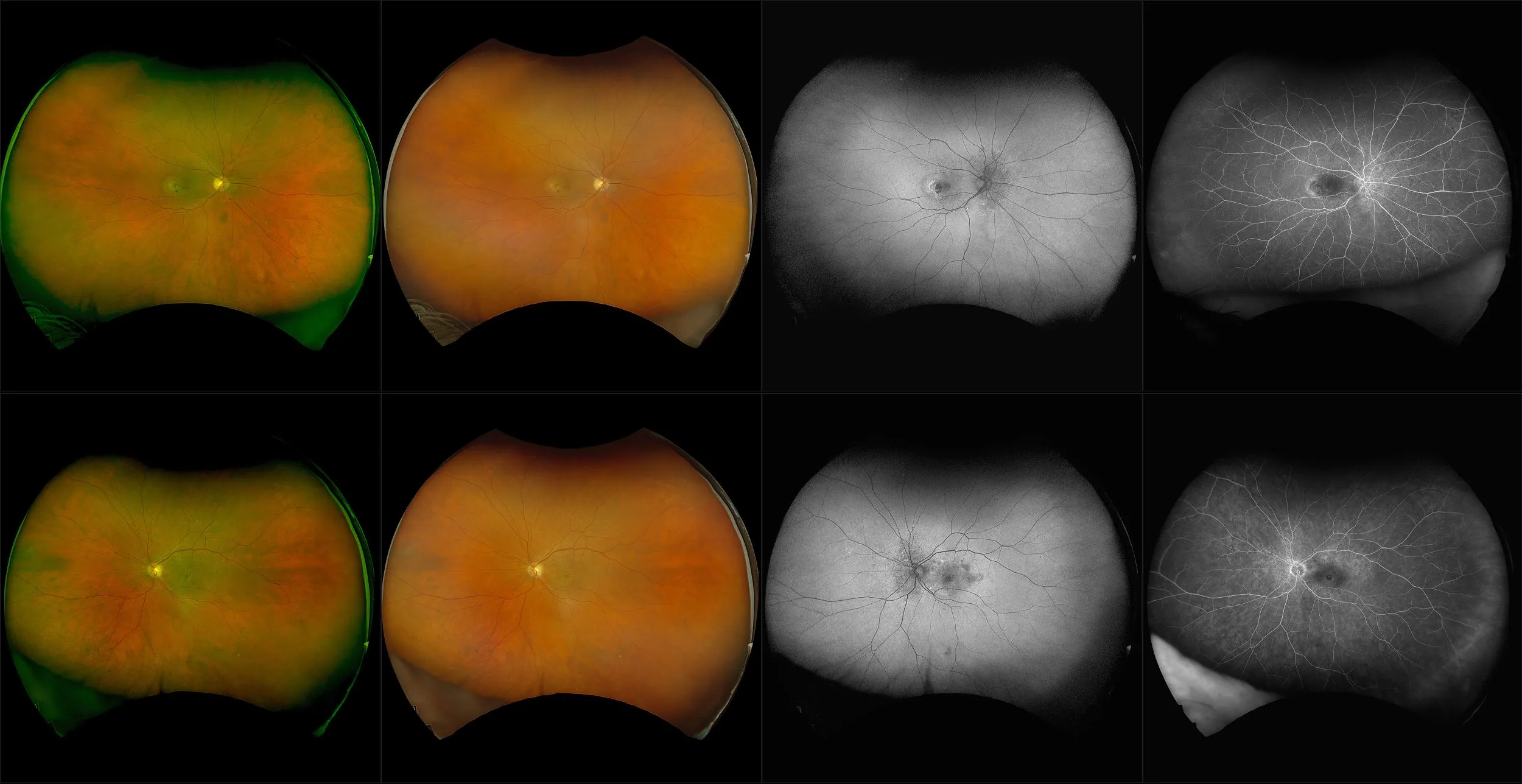
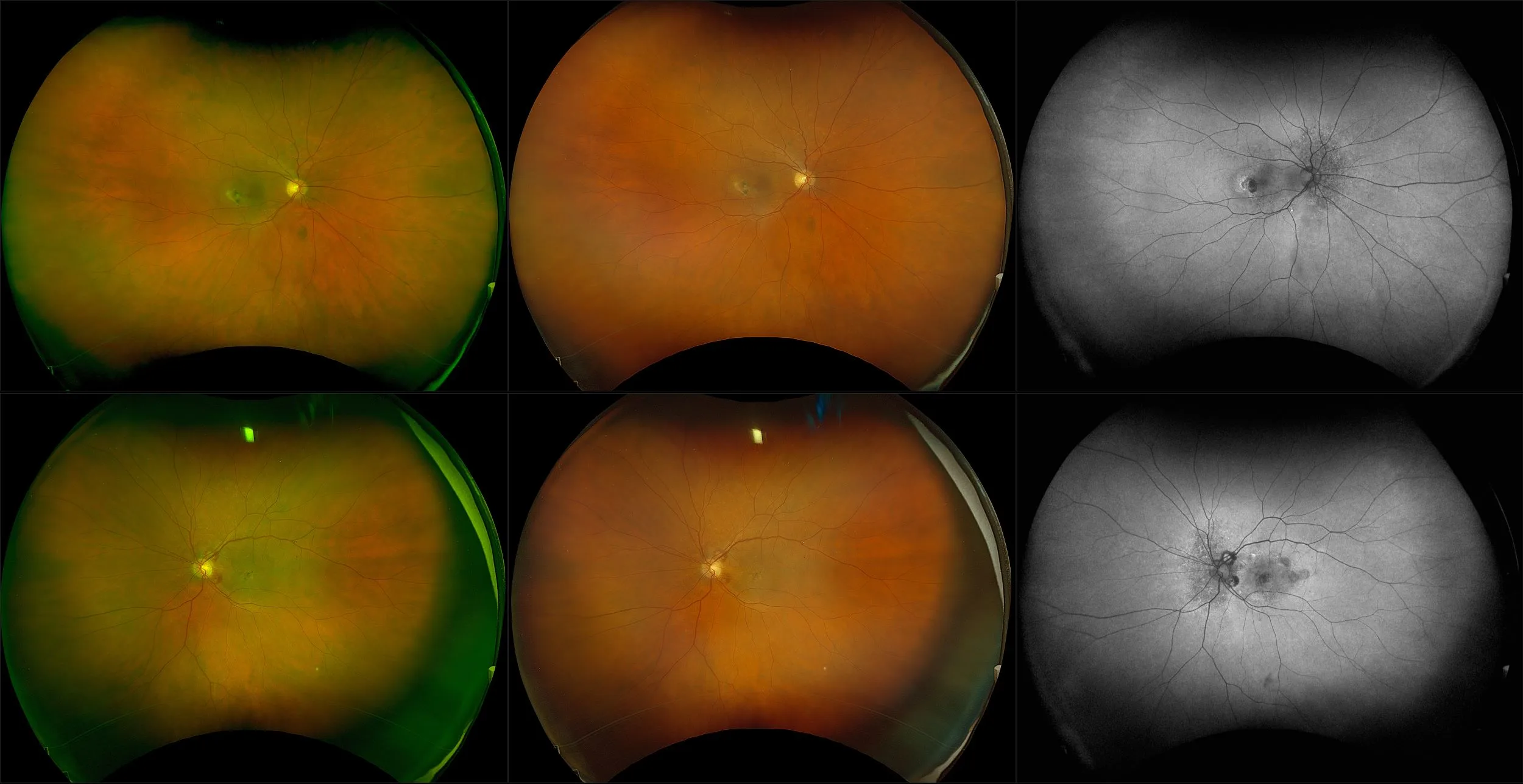
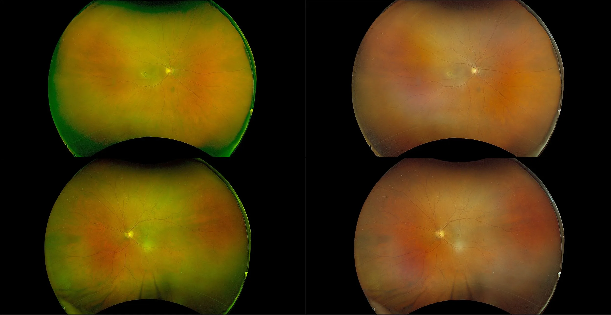
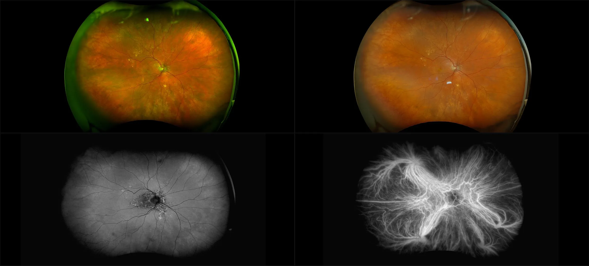
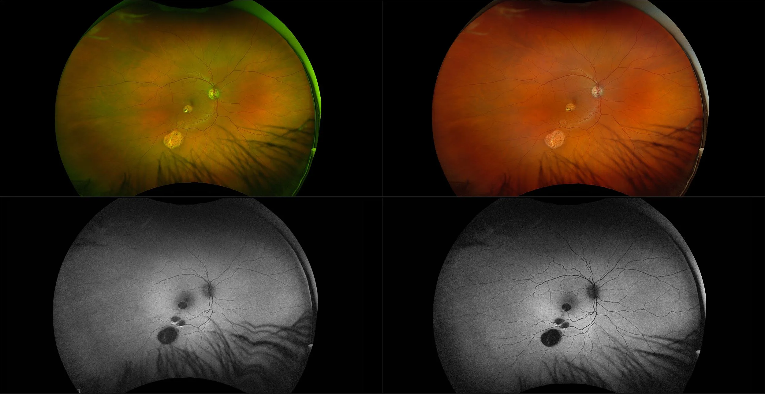
Choroidal Disease
The choroid is the pigmented, highly vascular layer of the globe, lying between the sclera (on the outside) and the retina (on the inside). It is one of the three components of the uveal tract and is shaped a little like the body of a rounded wine glass. The optic nerve emerges at its base and the other two components of the uveal tract sit anteriorly (the ciliary body lies around the rim and the iris stretches over the opening). It is made up of three layers, each of which can be affected by disease processes. There is the external vessel layer, the capillary layer and the internal sheet-like Bruch's membrane. The choroid can be subject to inflammatory disorders (often in conjunction with the retina - see the separate Chorioretinal Inflammation article), tumor formation, and a number of other disorders. https://patient.info/doctor/choroidal-disorders