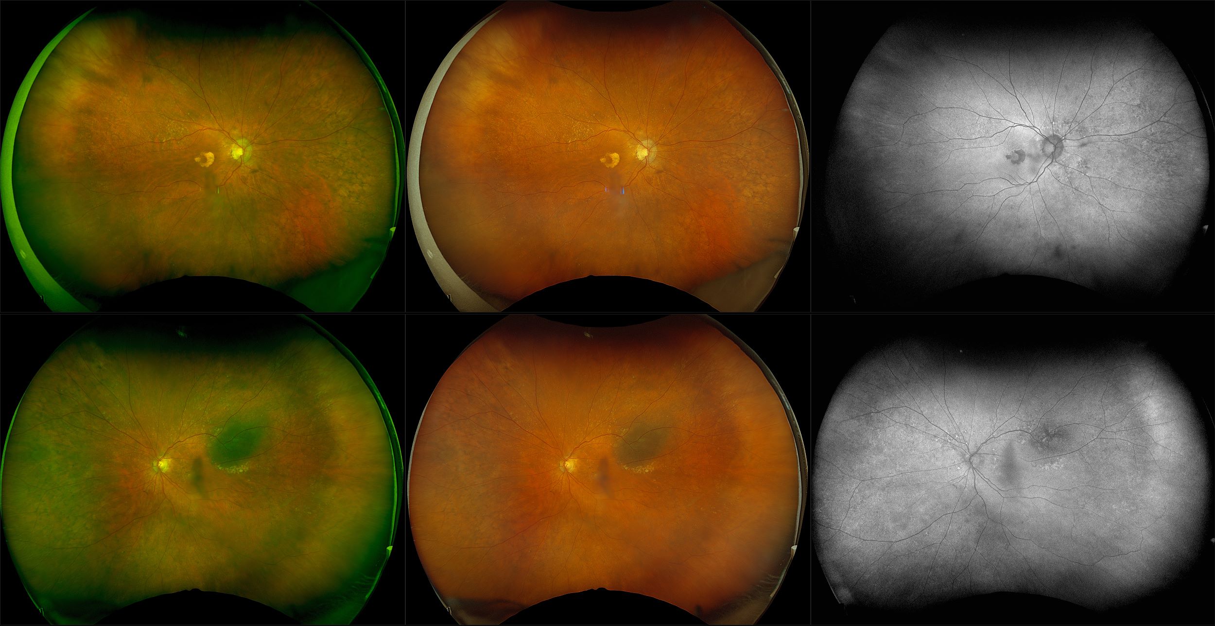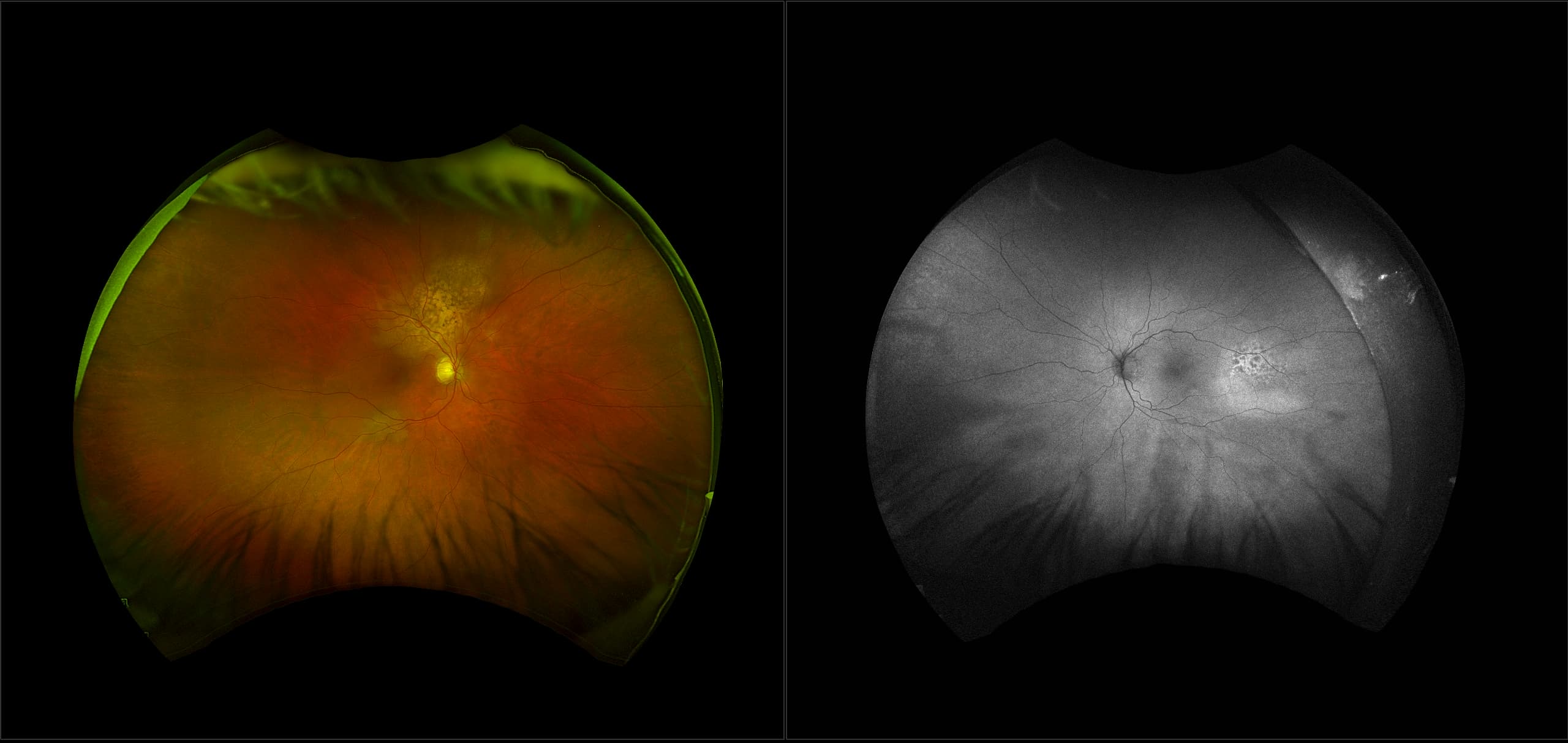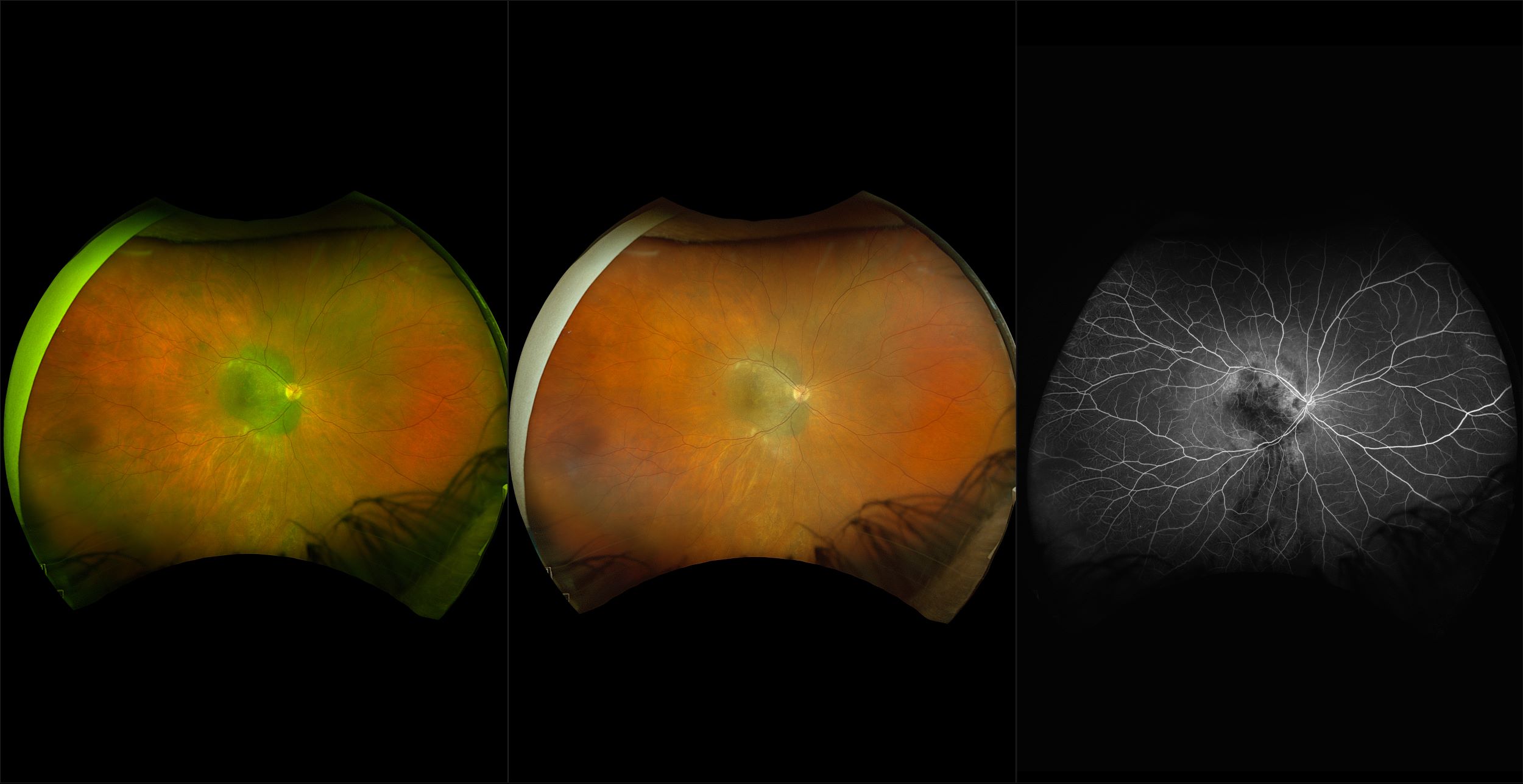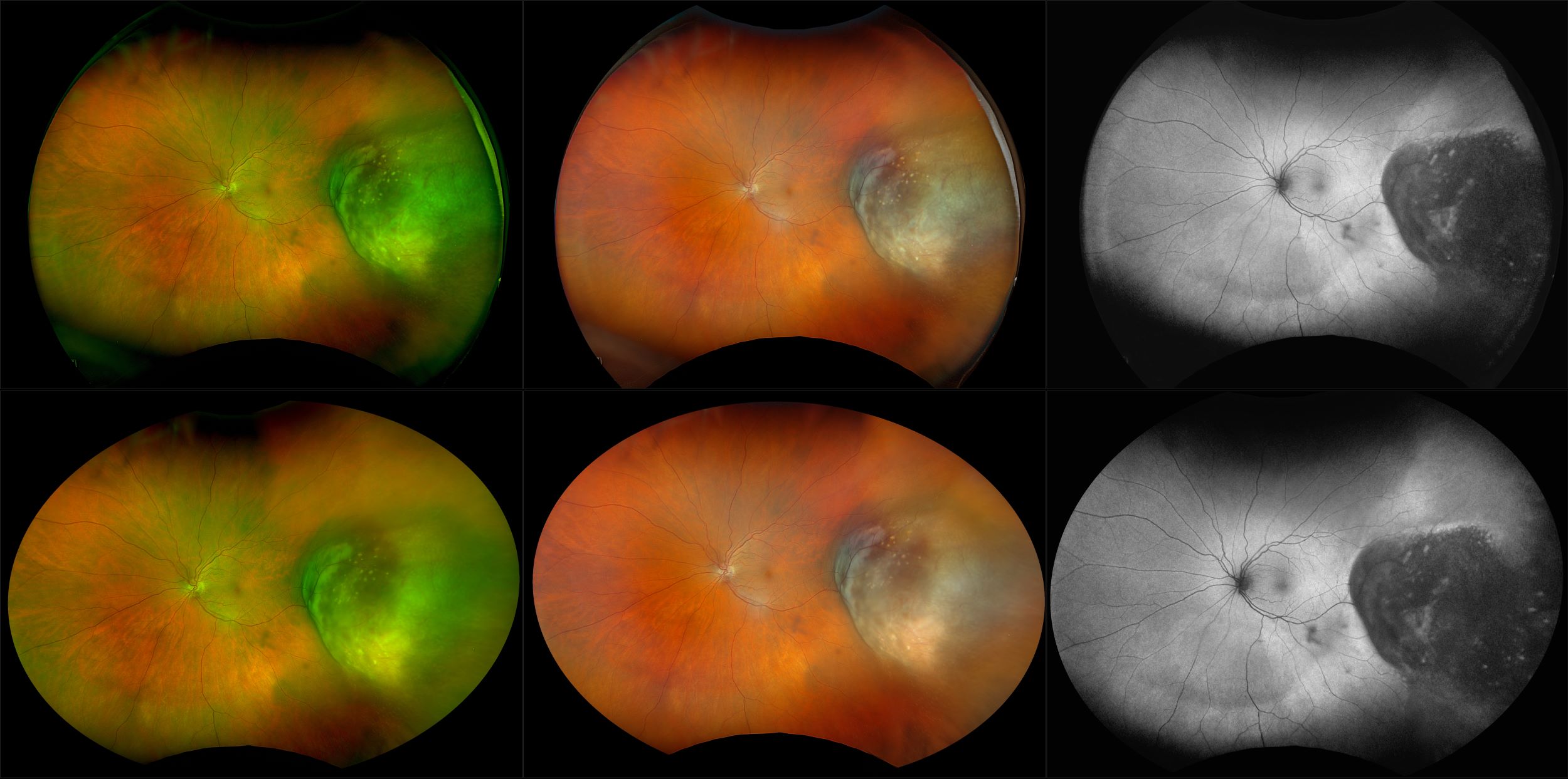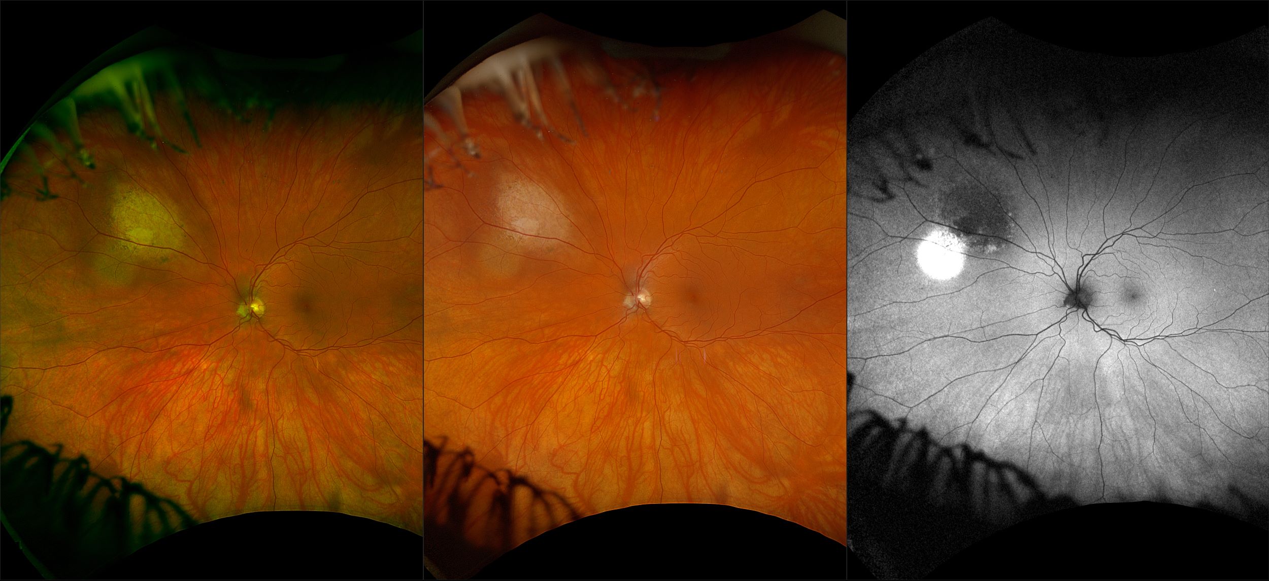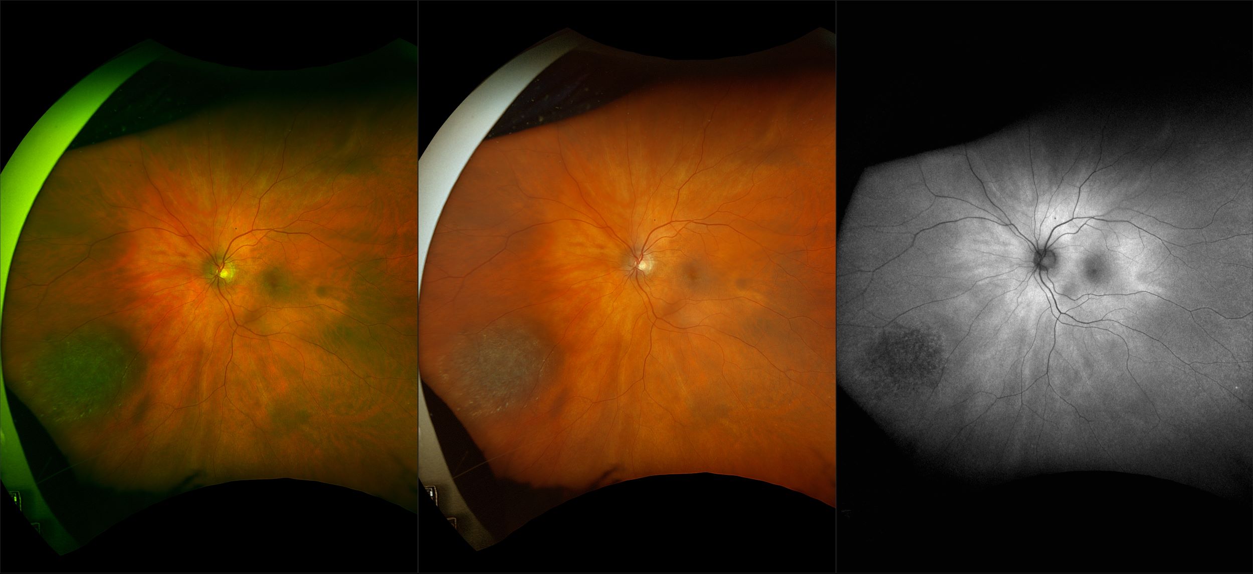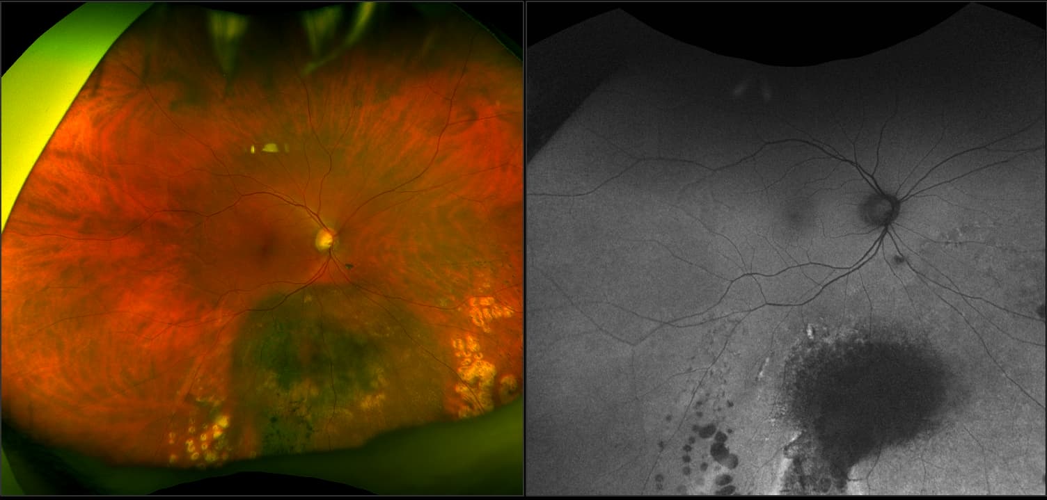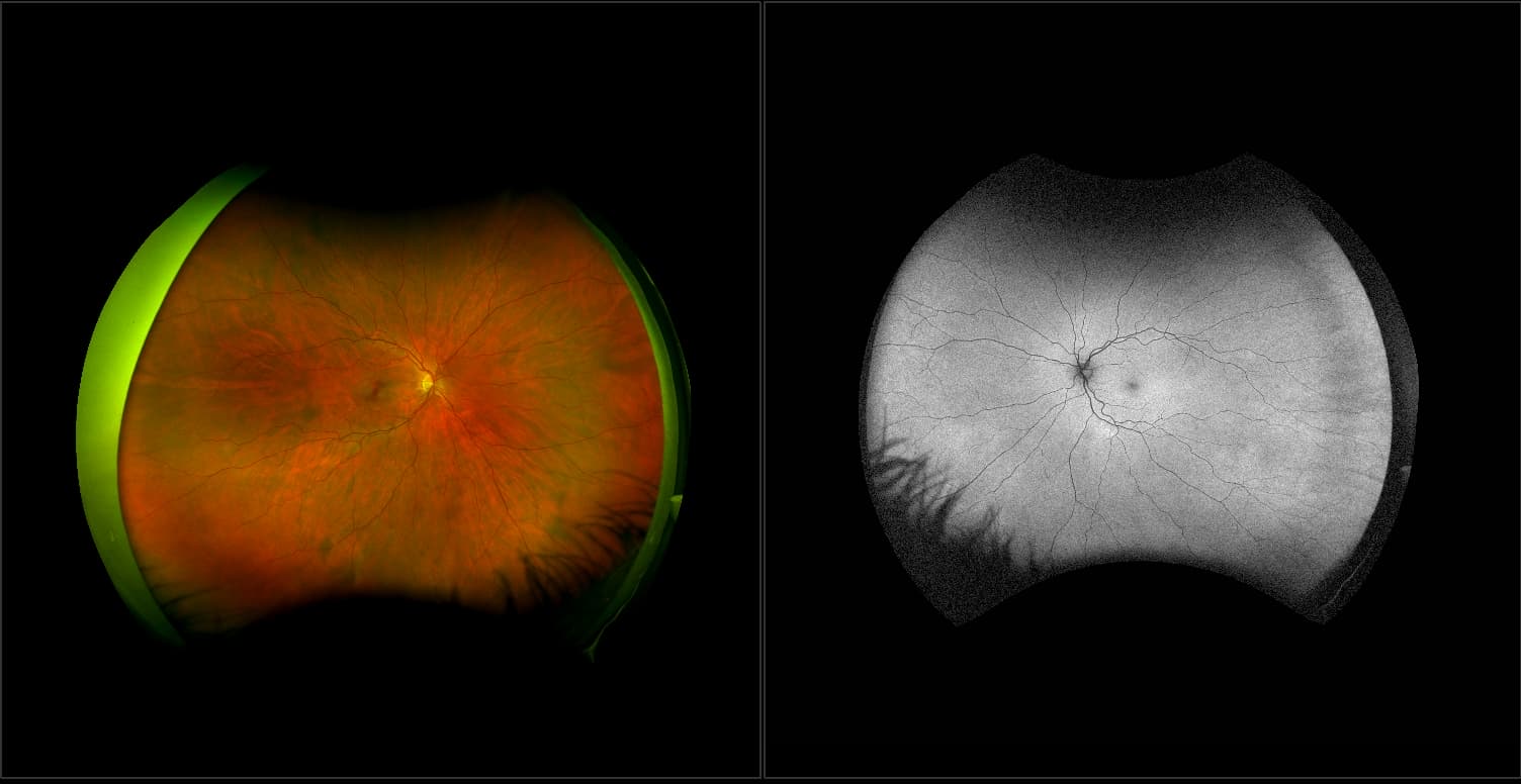
California - Choroidal Nevus, RG, AF
What: Choroidal nevi are flat, benign pigmented areas that appear in the back of the eye.
Why: Studies have shown the benefit of imaging choroidal nevi using a widefield scanning laser ophthalmoscope in that the two imaging channels (red 635nm and green 532nm) can be used to help determine the presence of choroidal nevi. Utilizing color optomap imaging increased the prevalence of visualizing choroidal nevi compared to other population-based studies where an ultra-widefield image was not used.
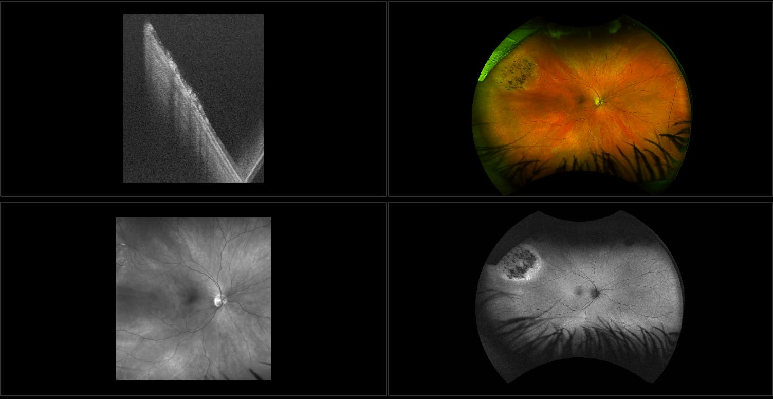
In the picture: optomap color rg from Dr. Paulo Stanga, Manchester Royal Eye Center, shows 3 choroidal nevi. When separated by the different channels, the nevi will disappear in the green channel and appear in the red channel showing the depth and location of the nevi in the choroid.
References:
- Gordon-Shaag A , Barnard S, Millodot M, Gantz L, Chiche G, Vanessa E, Ruth W, Pinchasov R, Gosman
Z, Simchi M, Koslowe K & Shneor E. Prevalence of choroidal naevi using scanning laser ophthalmoscope.
Ophthalmic Physiol Opt 2014, 34, 94–101.