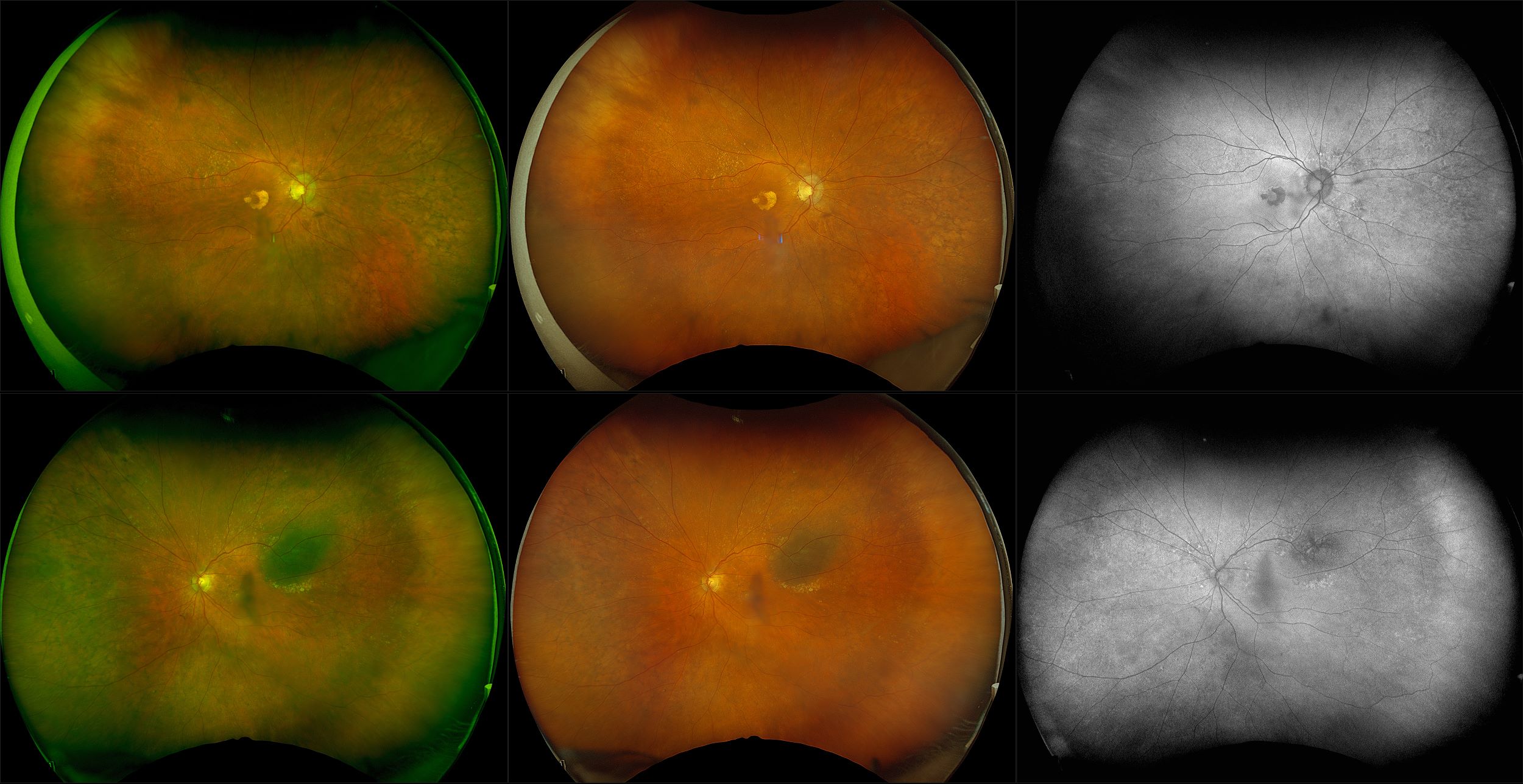
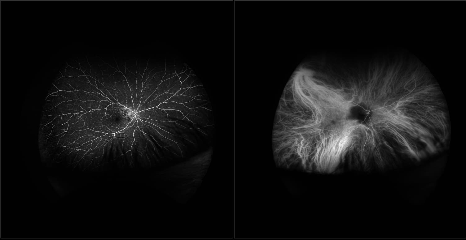
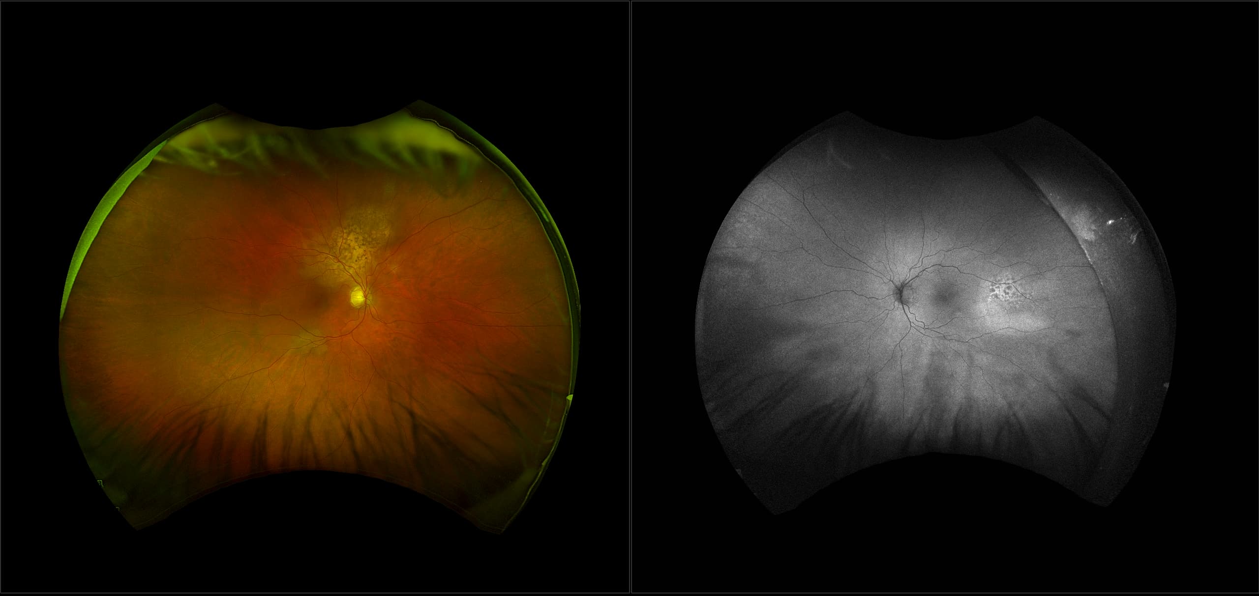
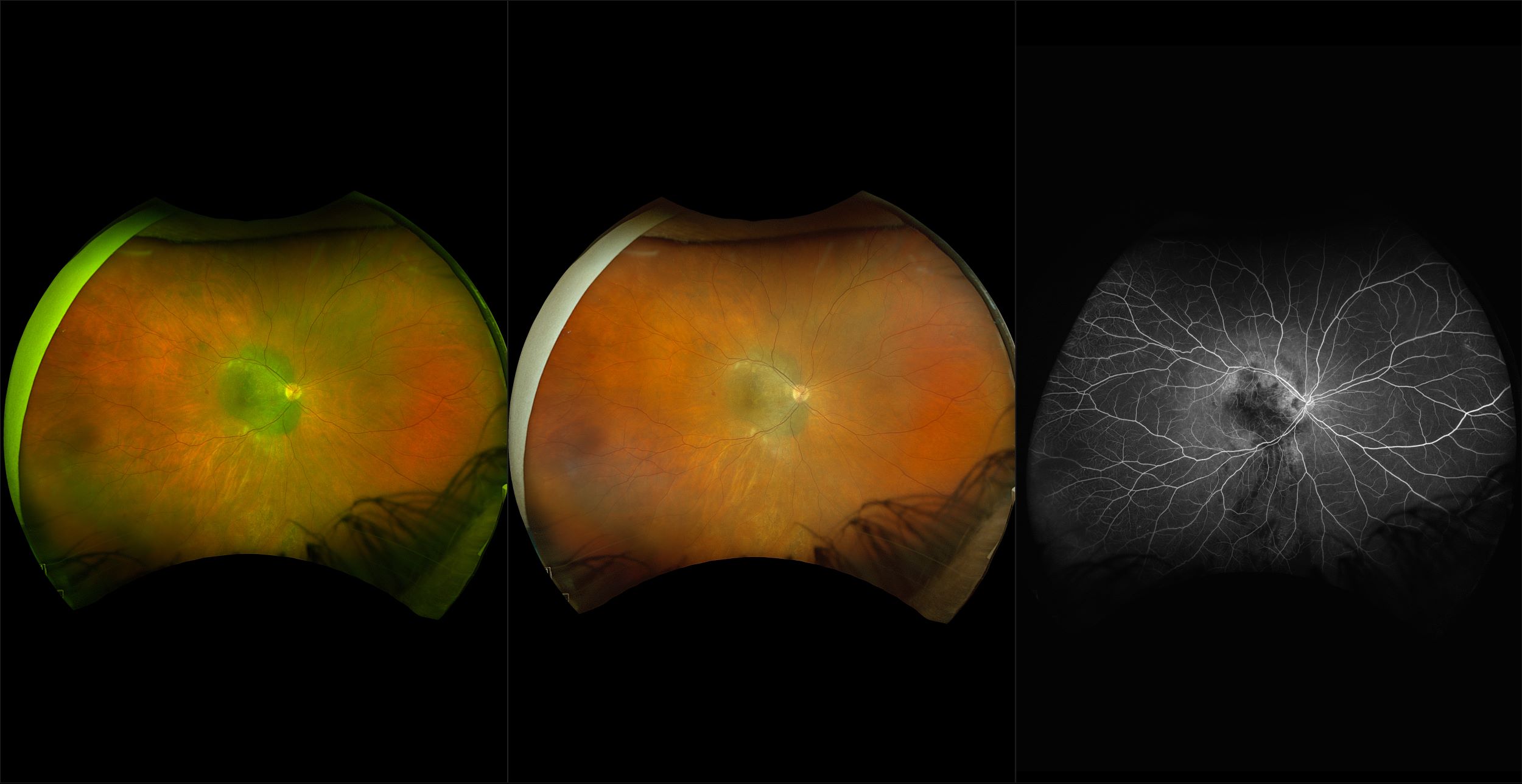
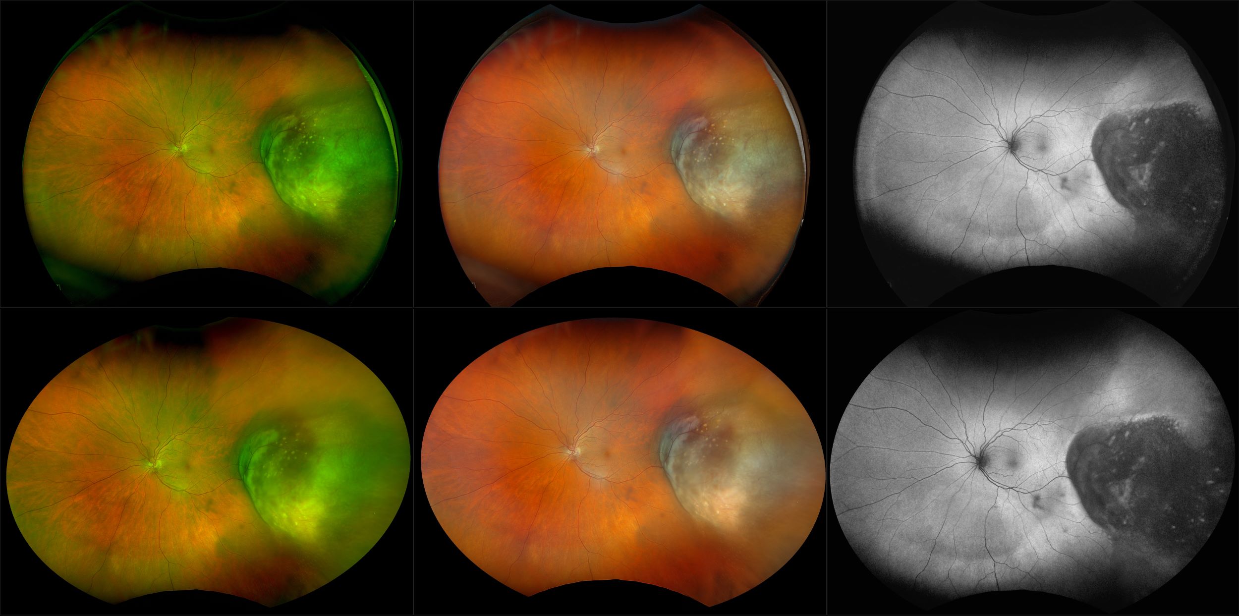
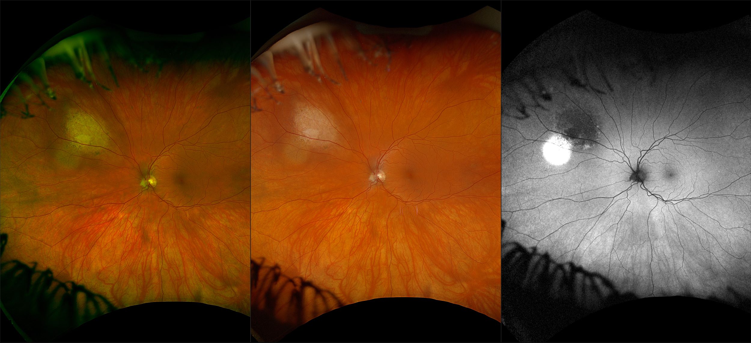
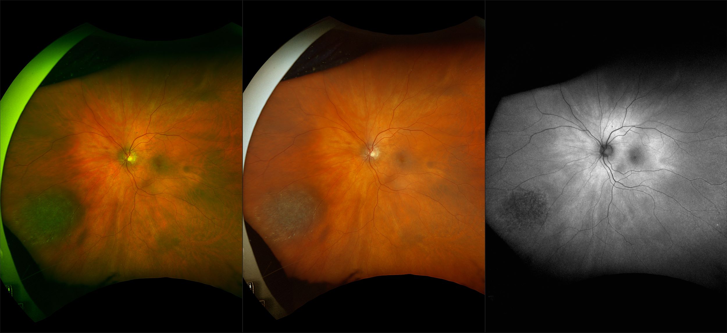
You are now leaving the Global site.

Ok Cancel
optos
  • Products
  • Patients
  • Providers
  • About Us
  • Contact
  • Customers
  • Language Menu
    English Deutsch Français
Home > Recognizing Pathology >

Choriodal Nevus

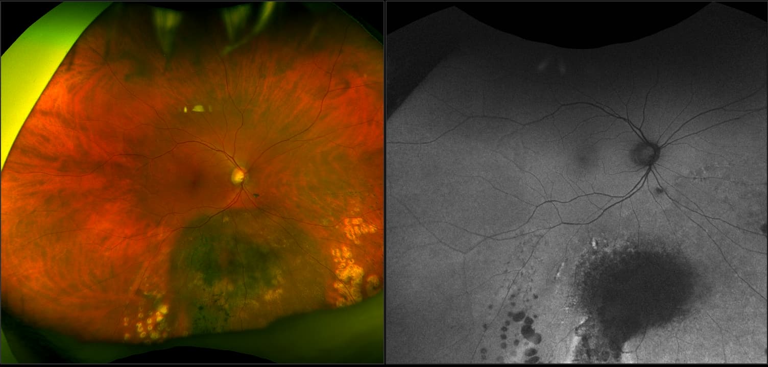
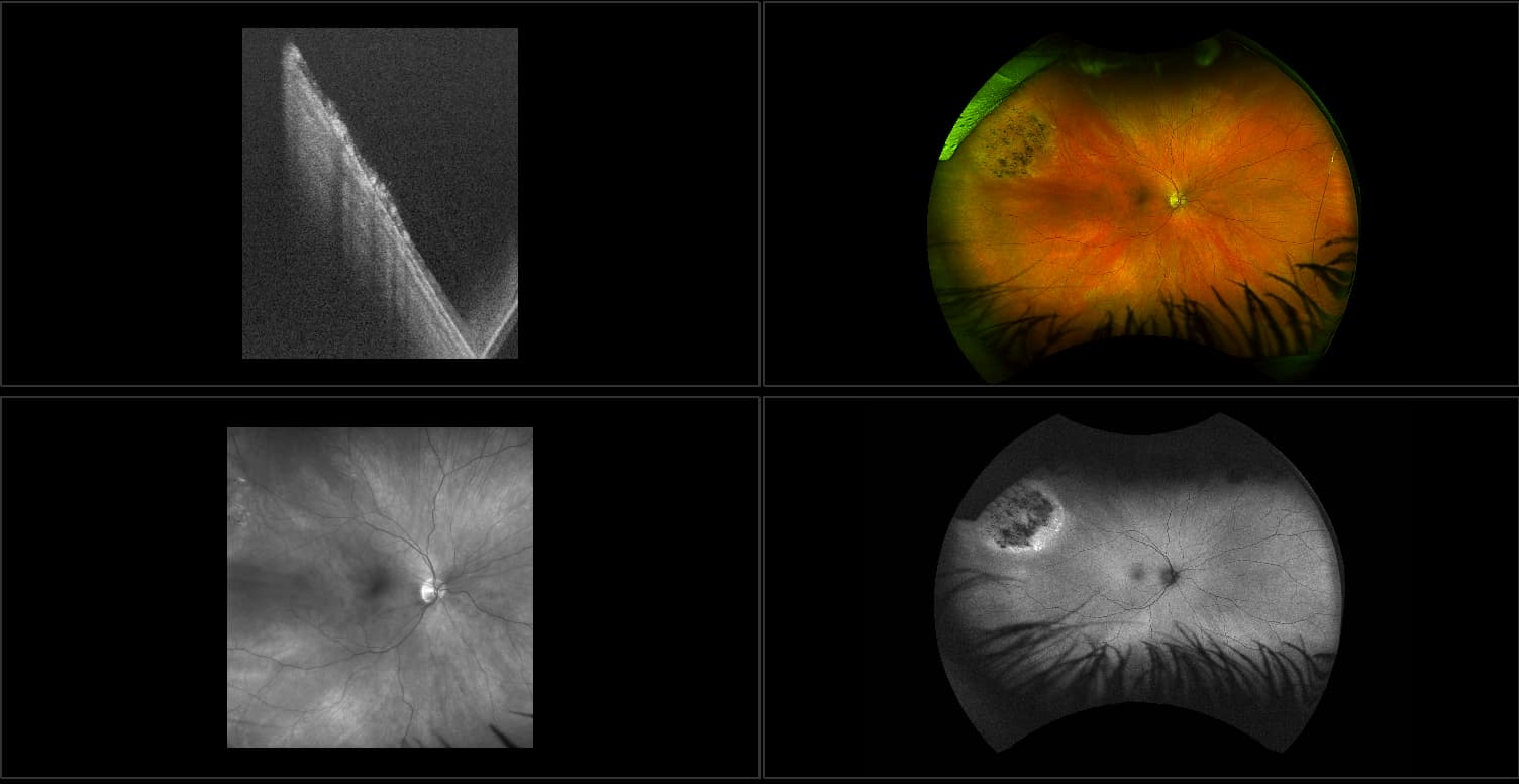
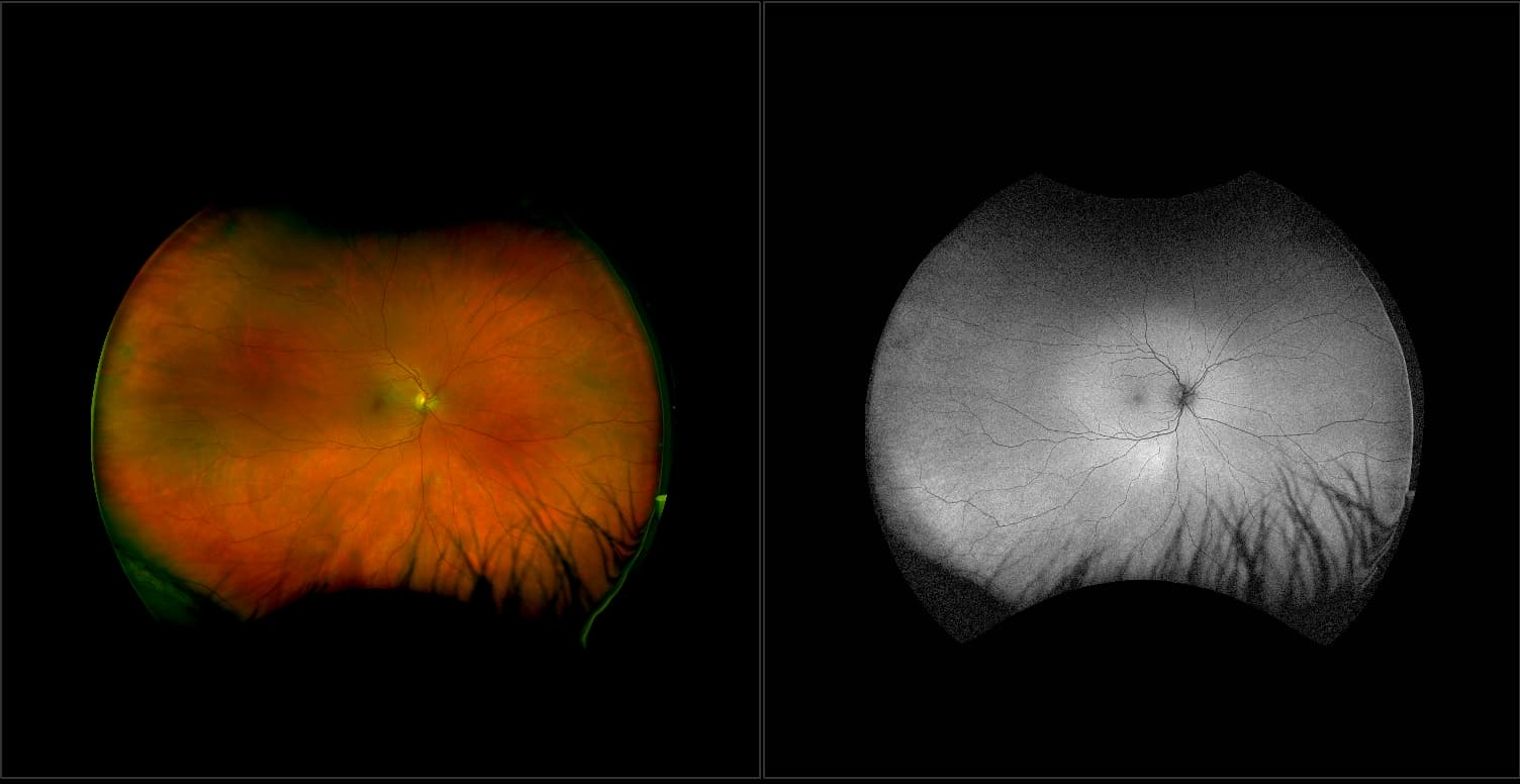
A choroidal nevus is simply an accumulation of melanocytes in the choroid. They are a space occupying mass or a benign melanoma. Choroidal nevi are slate gray in color, have indistinct margins, and may be slightly elevated (1-2 diopters). Since they are in the choroid, they are usually only seen in the red separation and not the green. The green separation may show the RPE degeneration associated with nevi which is seen as pigment mottling and drusen.
View Full Case

Device

Image Modality

  • optomap af

How can we help?

If you have questions regarding the cases, images, or pathologies, or if you would like further information about our ultra-widefield retinal imaging technology, please complete the form below.
I want to receive email updates about Optos products and services
<p>You are now leaving the Global site.</p> Ok Cancel
  • Blog
  • Cookie Policy
  • Careers
  • Distributor Portal
  • Manage Preferences
  • Media
  • News
  • Privacy
  • Registered in Scotland
  • UK Modern Slavery Act

© 2023 Optos. All rights reserved. Company Registration Number SC139953