Age-related Macular Degeneration (AMD)
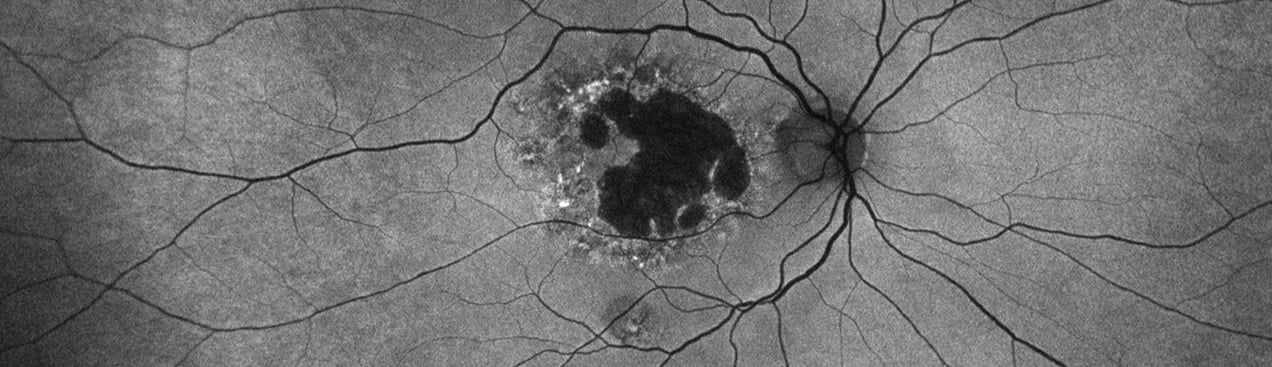
Age-related macular degeneration is found in two forms 1. Dry early form. 2. Wet (serous leakage or whole blood). Early dry form typically has drusen (>63u) and RPE degeneration and only needs periodic follow-ups, intermediate dry form has large drusen (>125u) and RPE degeneration and proliferation which requires examinations every 3 to 6 months depending on the severity of the presentation.
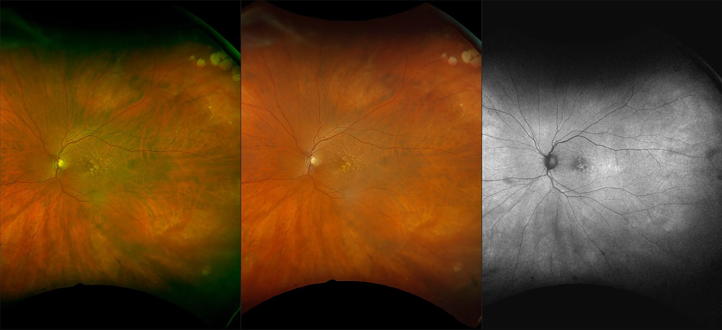
Studies have found that 97% of AMD patients have drusen in the periphery and concluded that “documentation of such finding may have implications regarding the risk of visual loss in AMD patients. AMD is NOT confined to the posterior pole” (Chew, et al. Peripheral Retinal changes associated with AMD in the Age-related eye disease study).
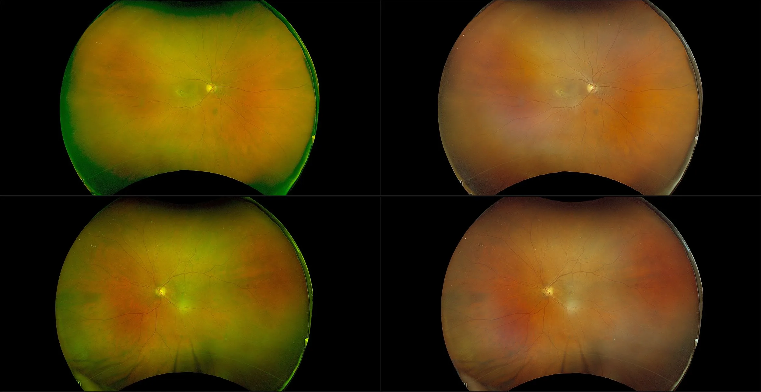
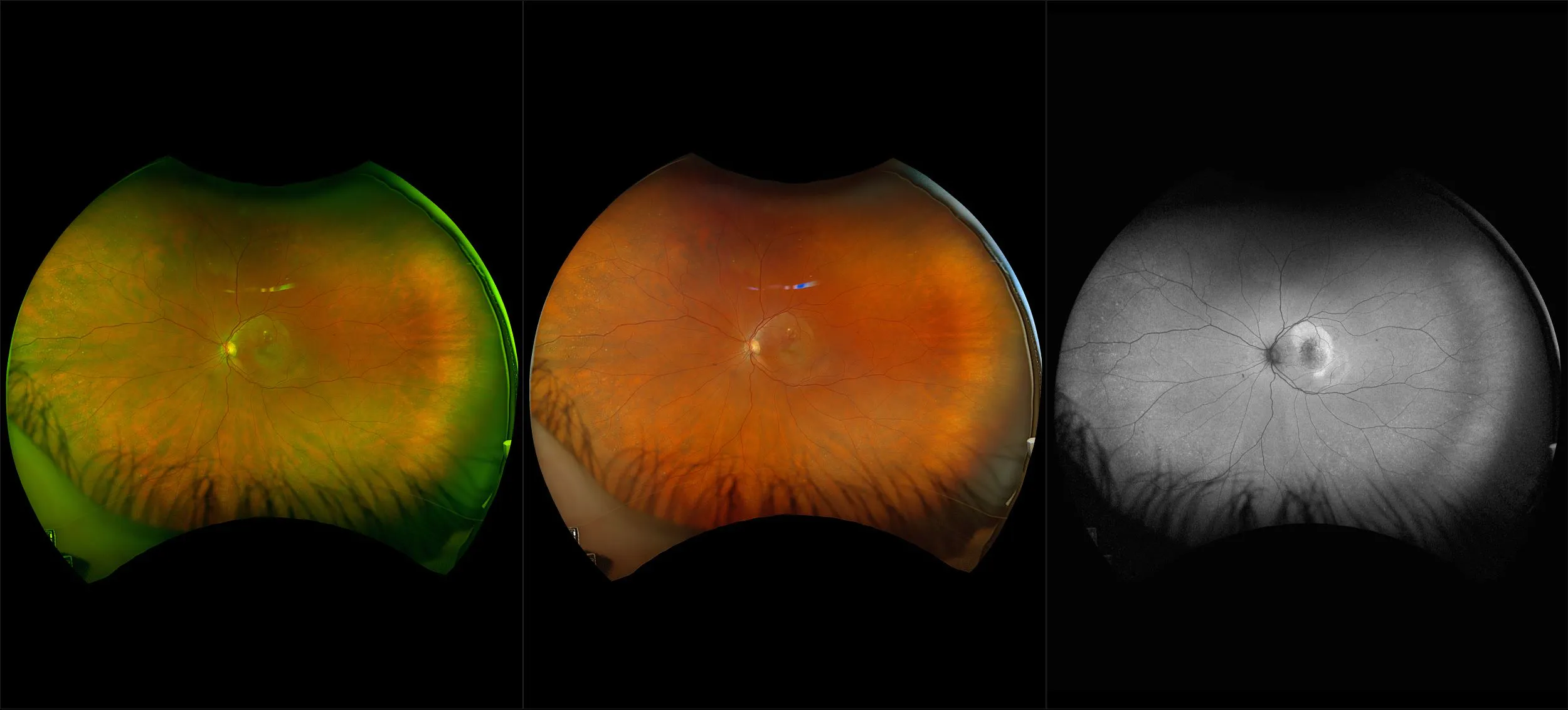
The recommended modality for utilizing optomap to visualize drusen, optomap Choroidal (the green channel) is captured with optomap color imaging.
Clinical Summary
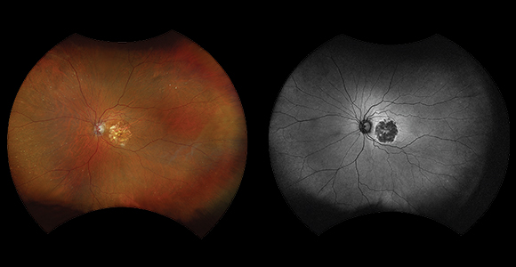
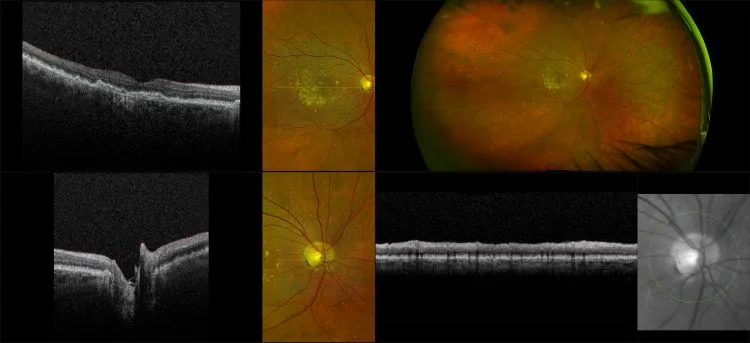
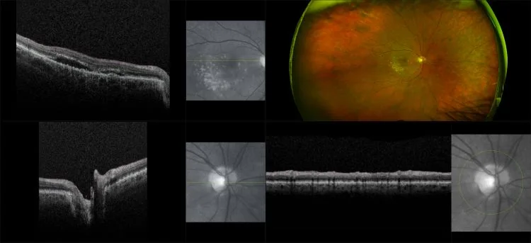
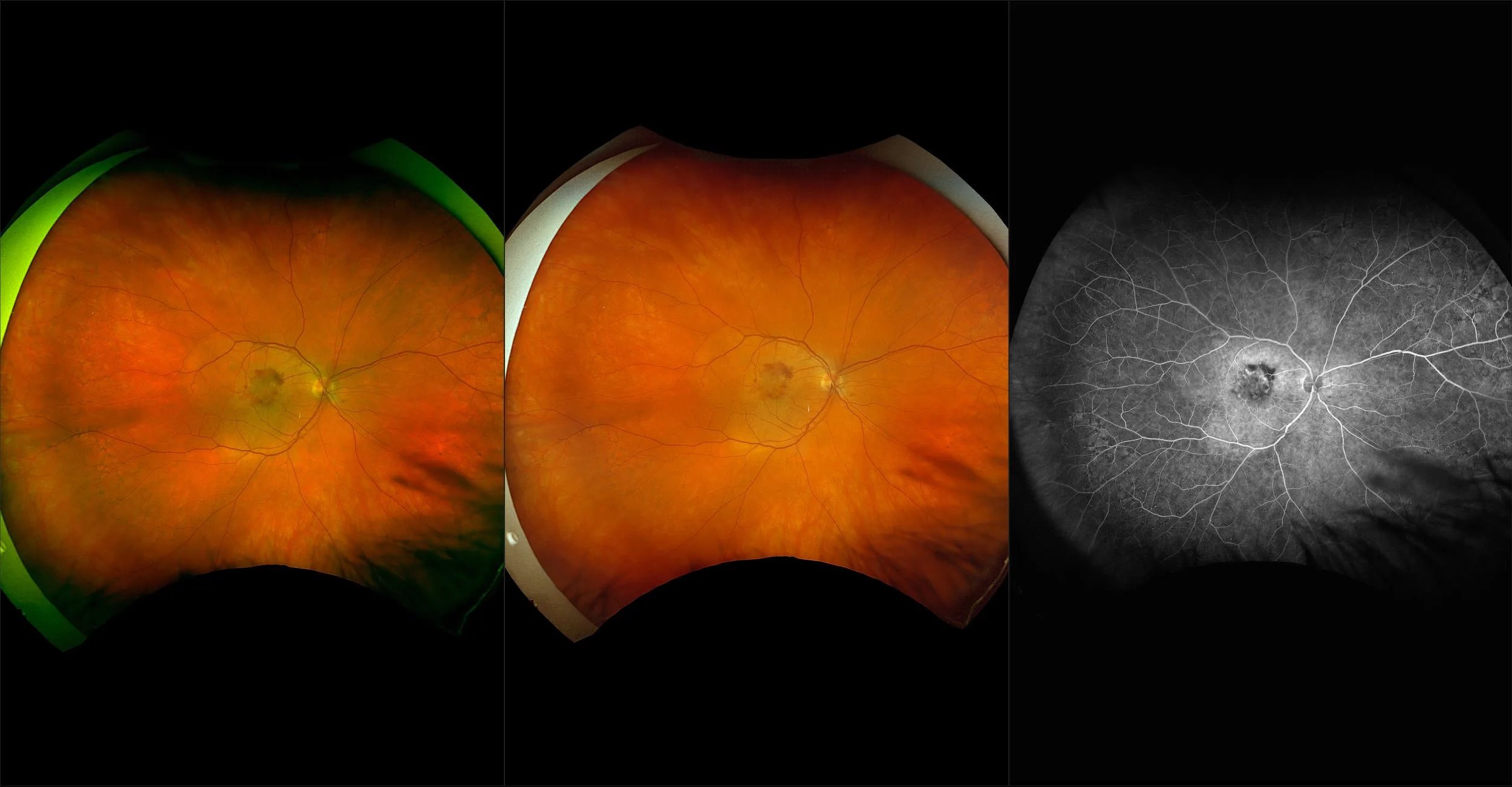
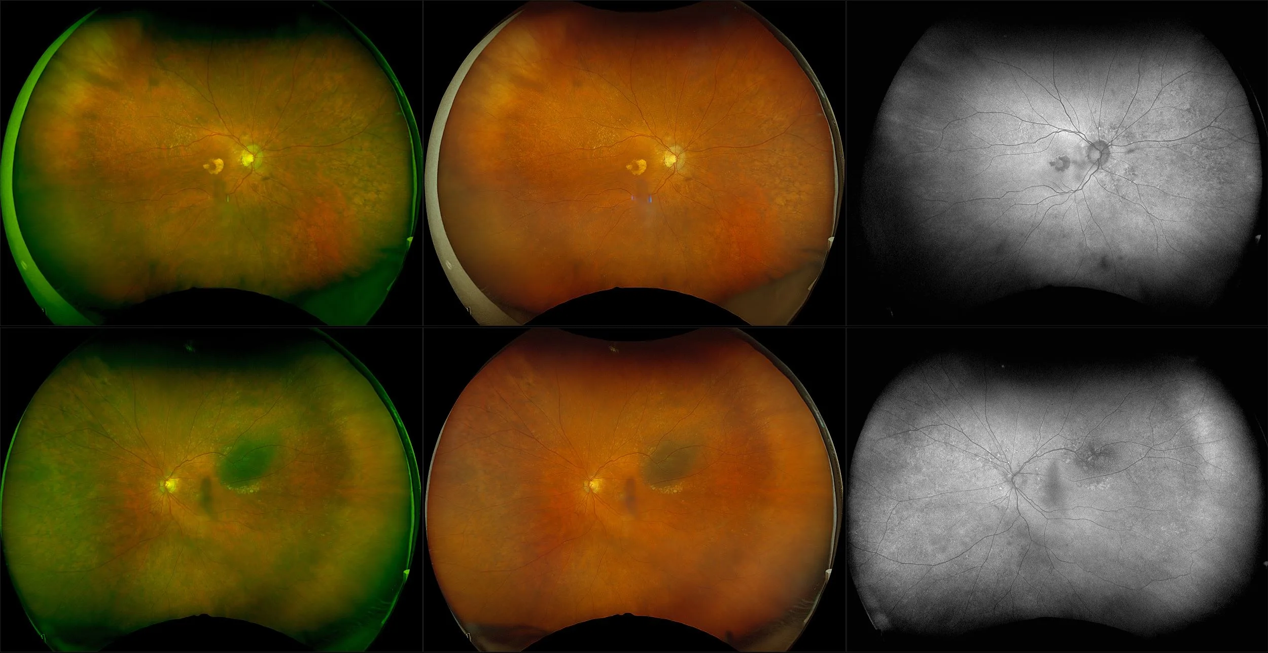
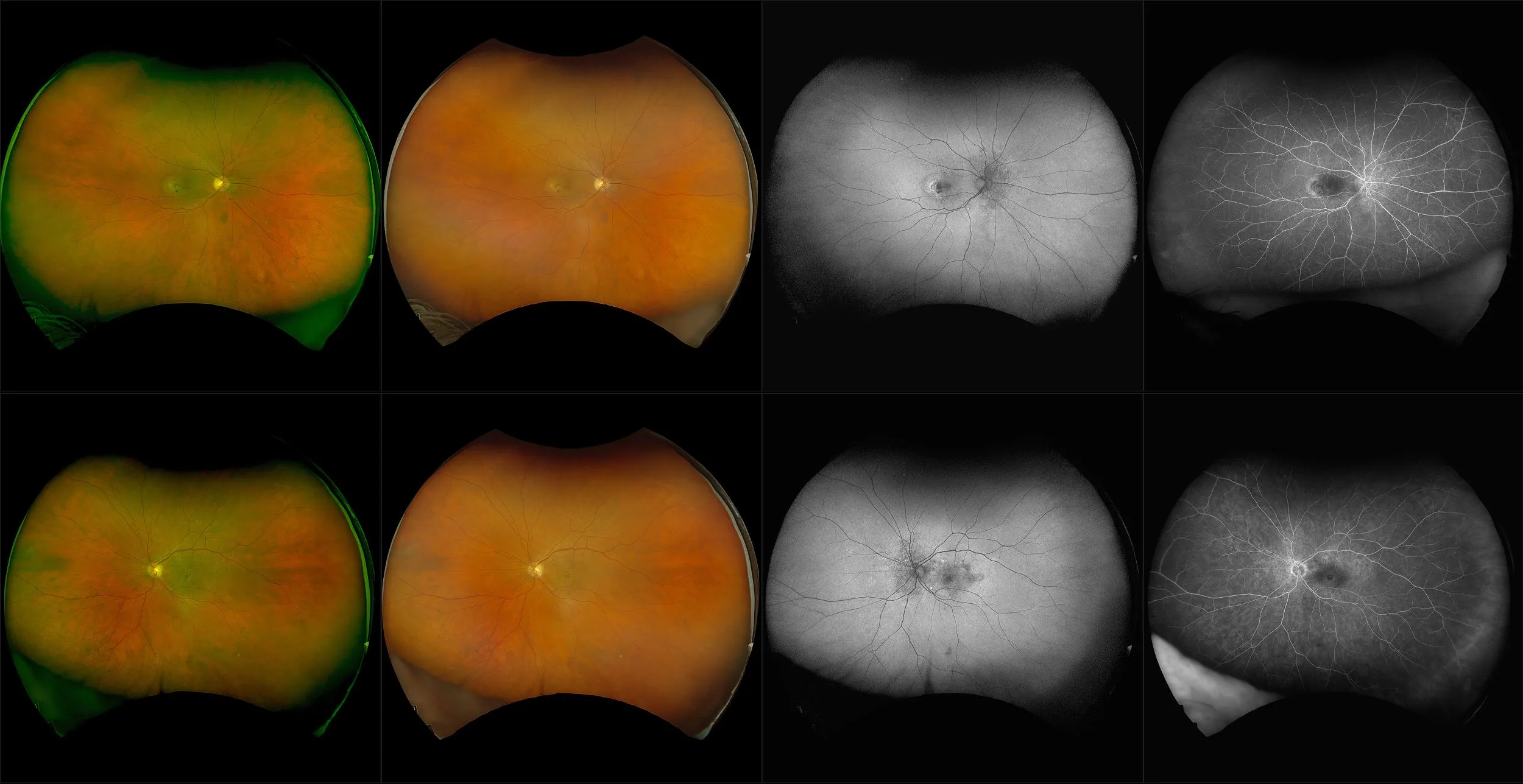
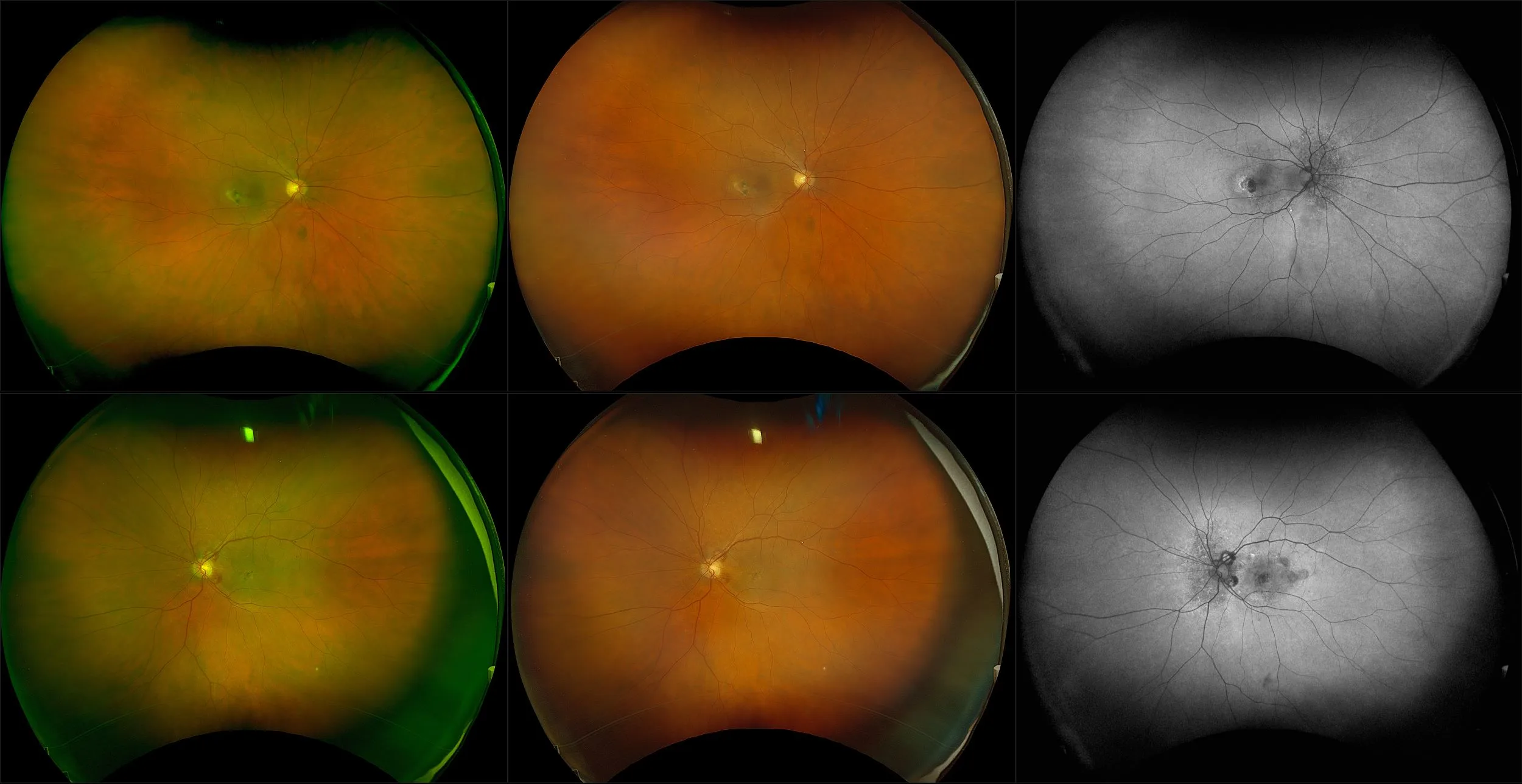
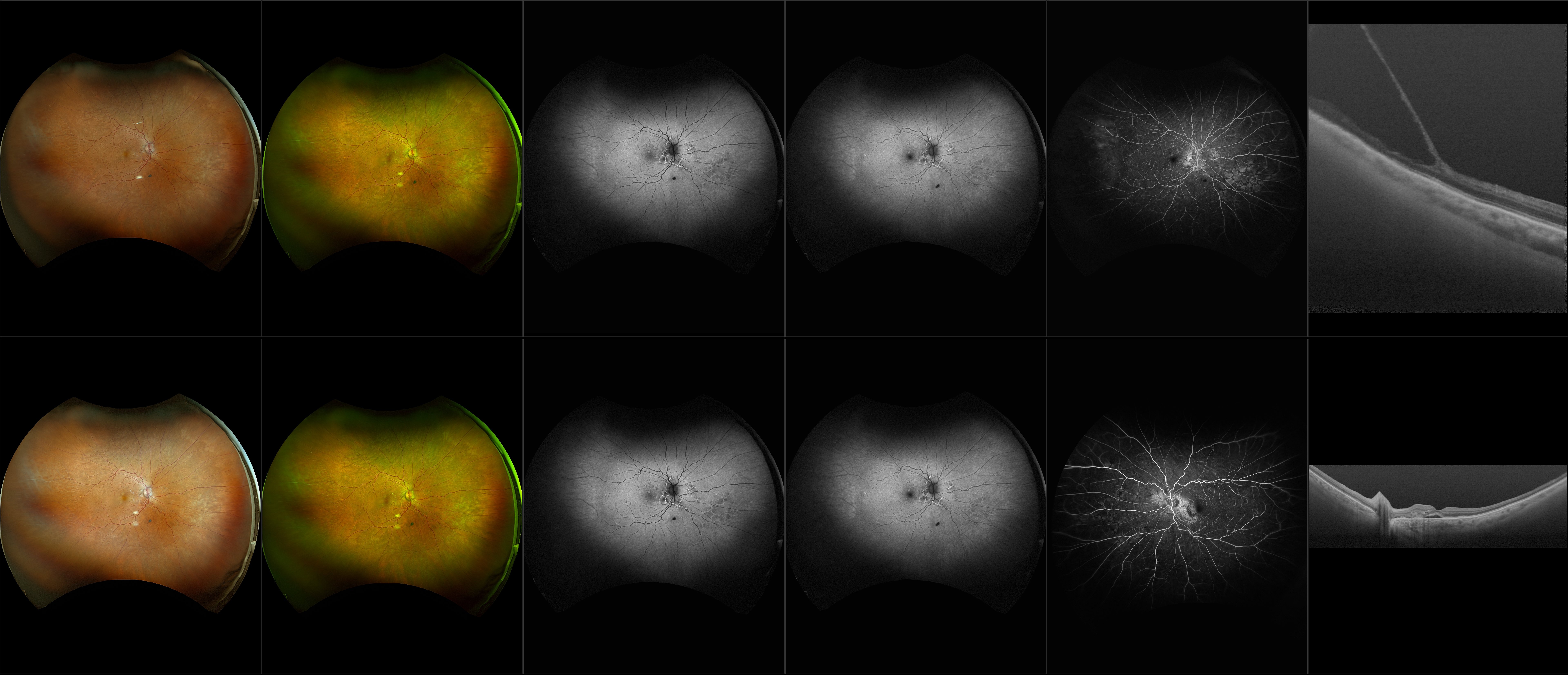
Multimodal optomap Images Enhance the Management of AMD
optomap has helped re-define AMD as a pan-retinal disorder.
Referenced Papers
Multimodal optomap Images Enhance the Management of AMD
optomap has helped re-define AMD as a pan-retinal disorder.
Evaluation of Peripheral Retinal Changes on Ultra-Widefield Fundus Autofluorescence Images of Patients with Age-Related Macular Degeneration.
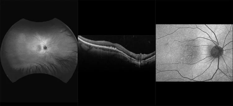
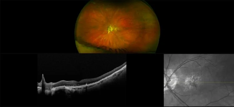
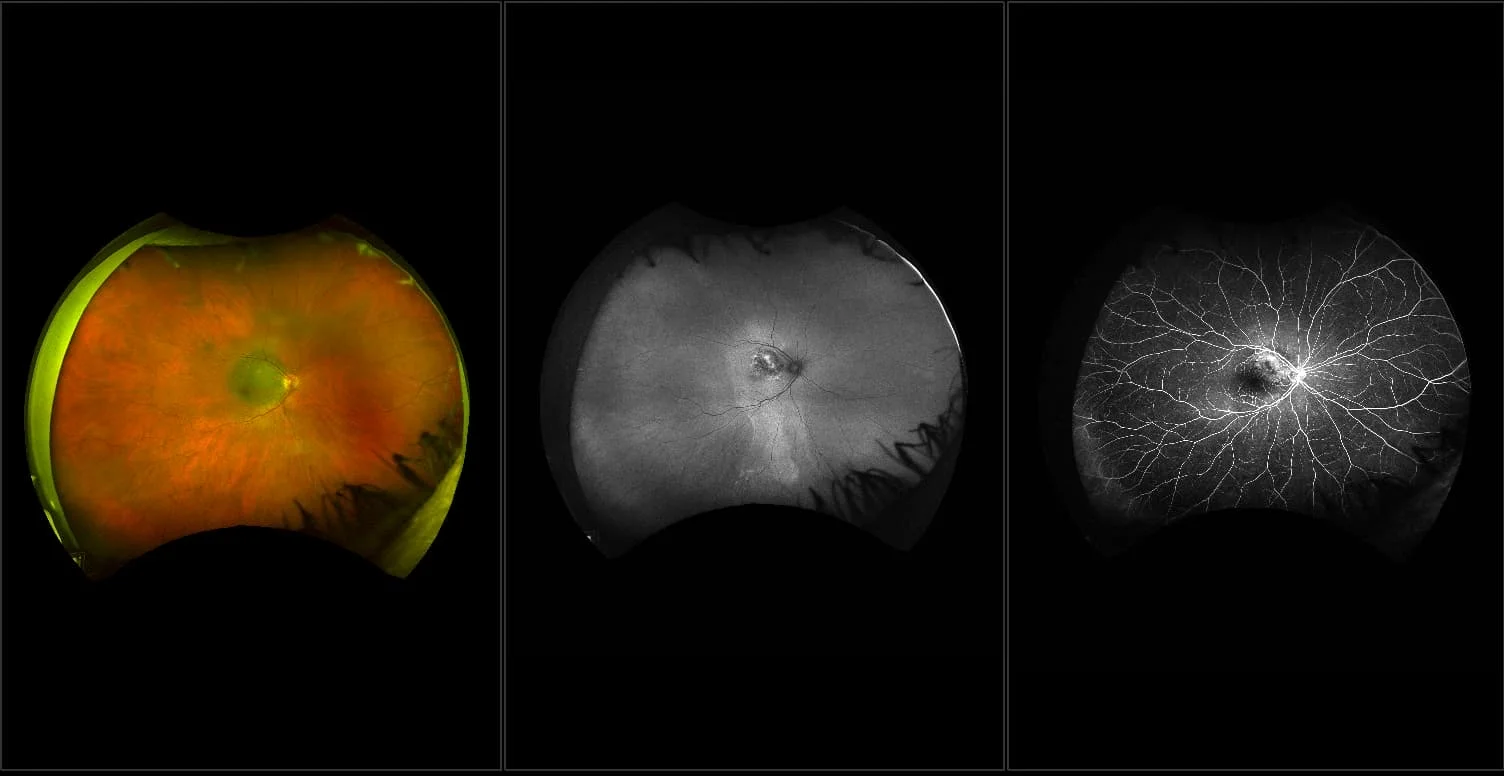
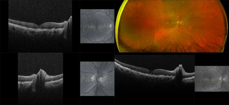
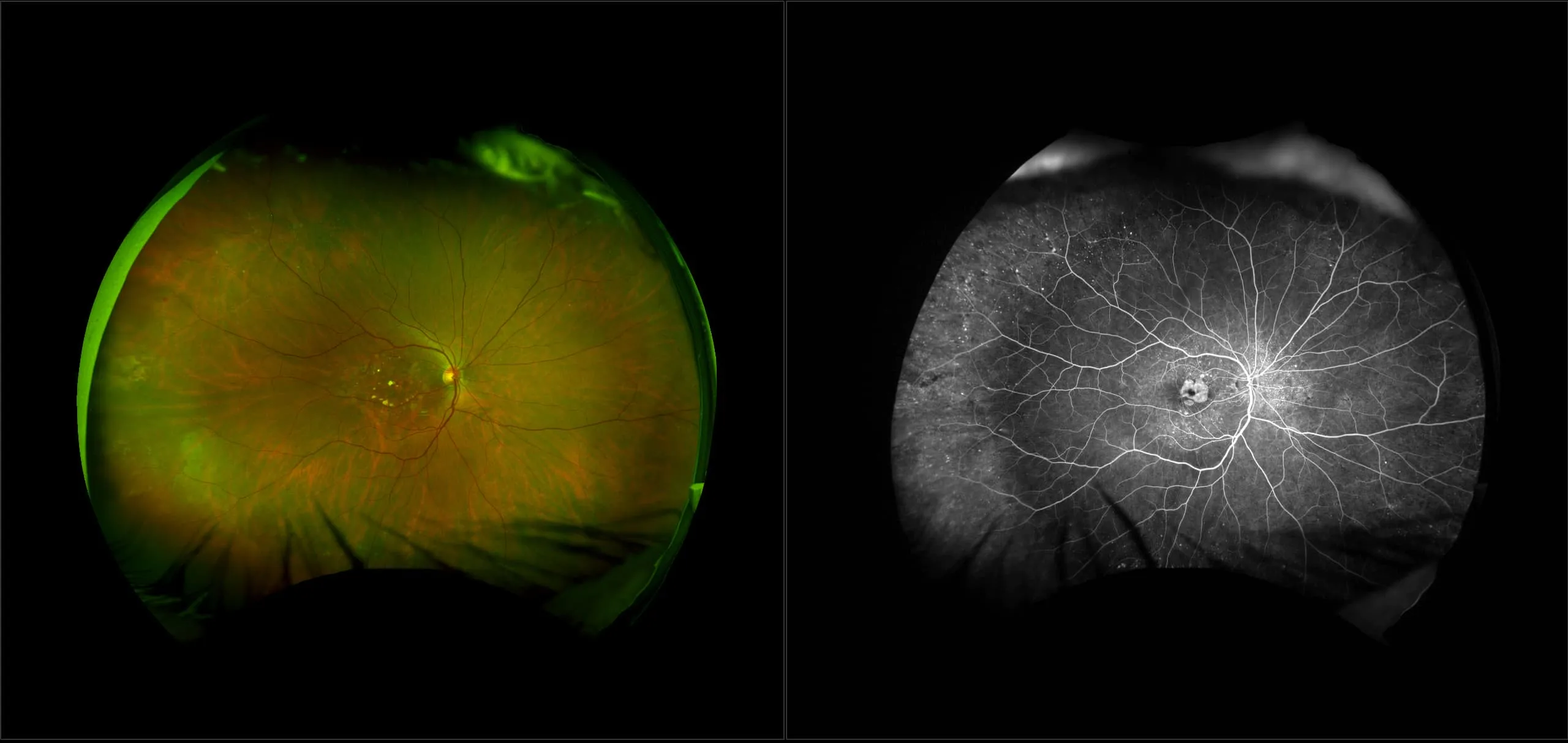
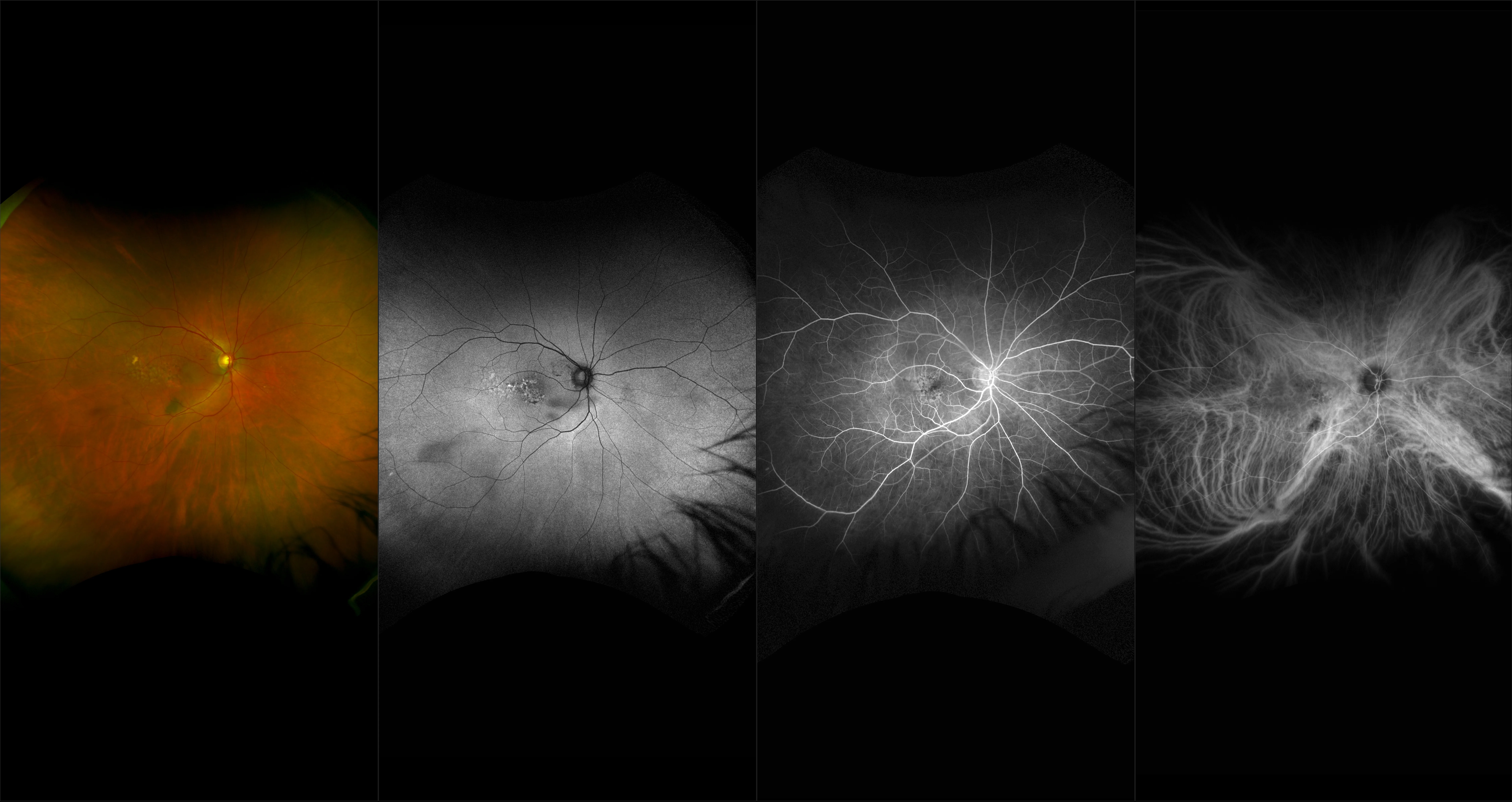
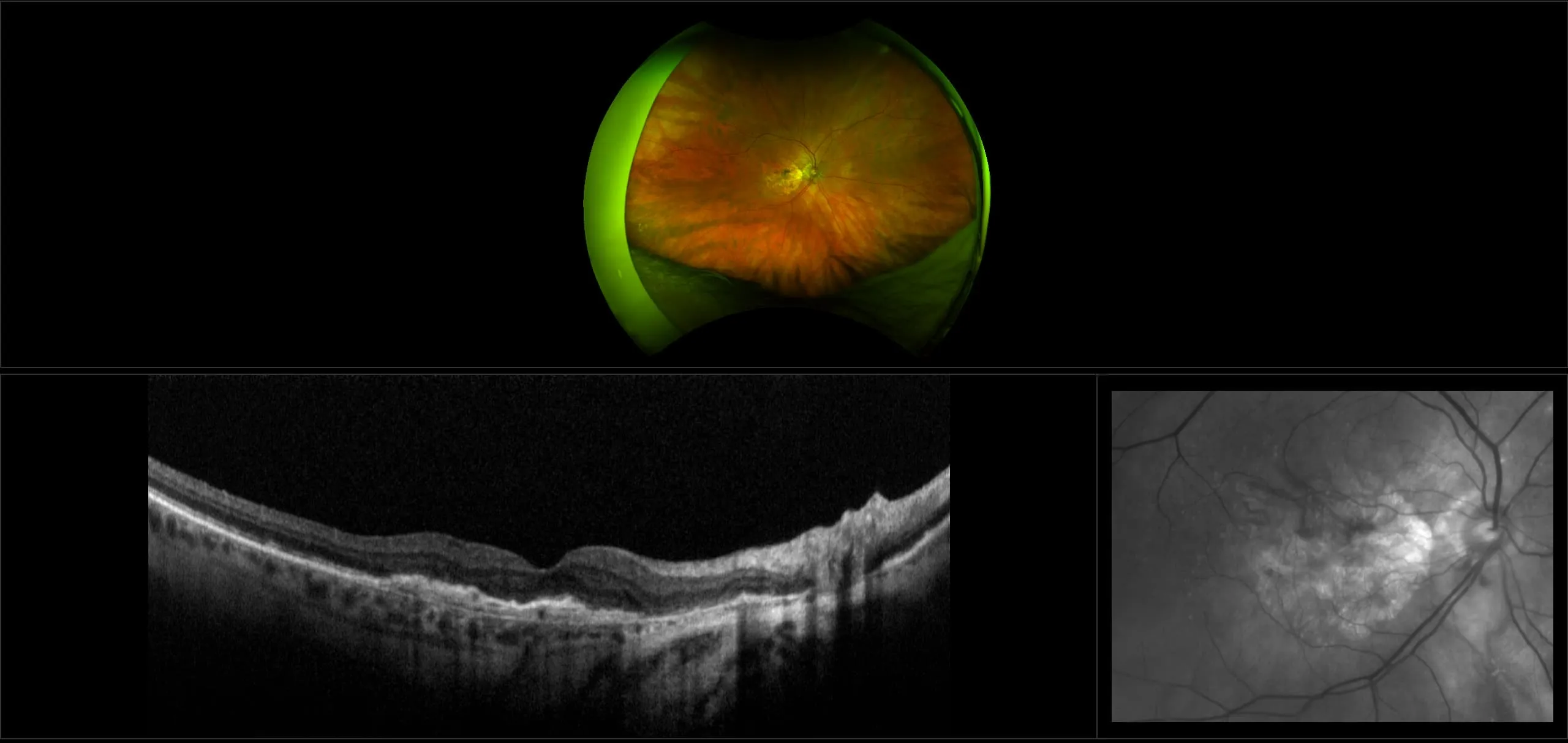
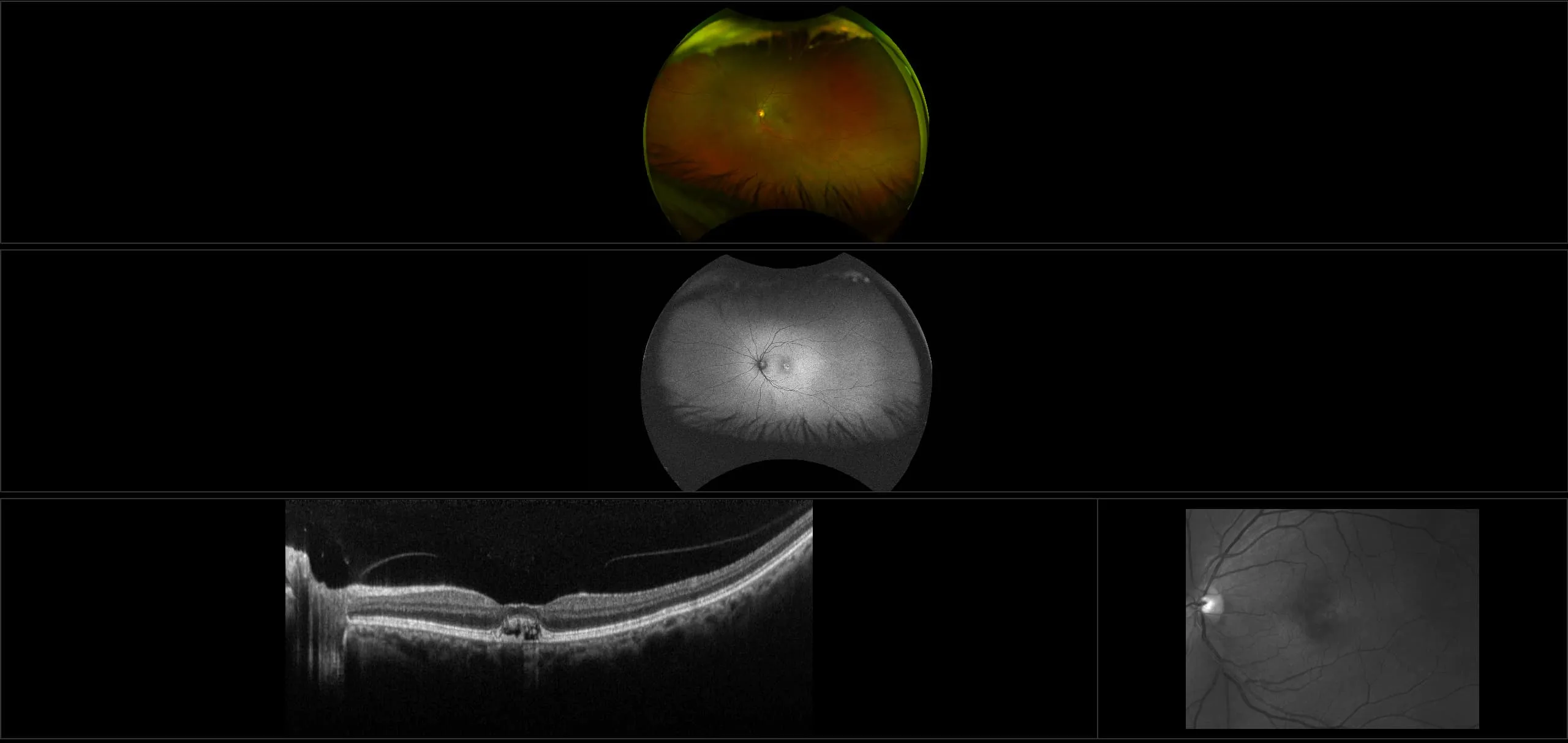
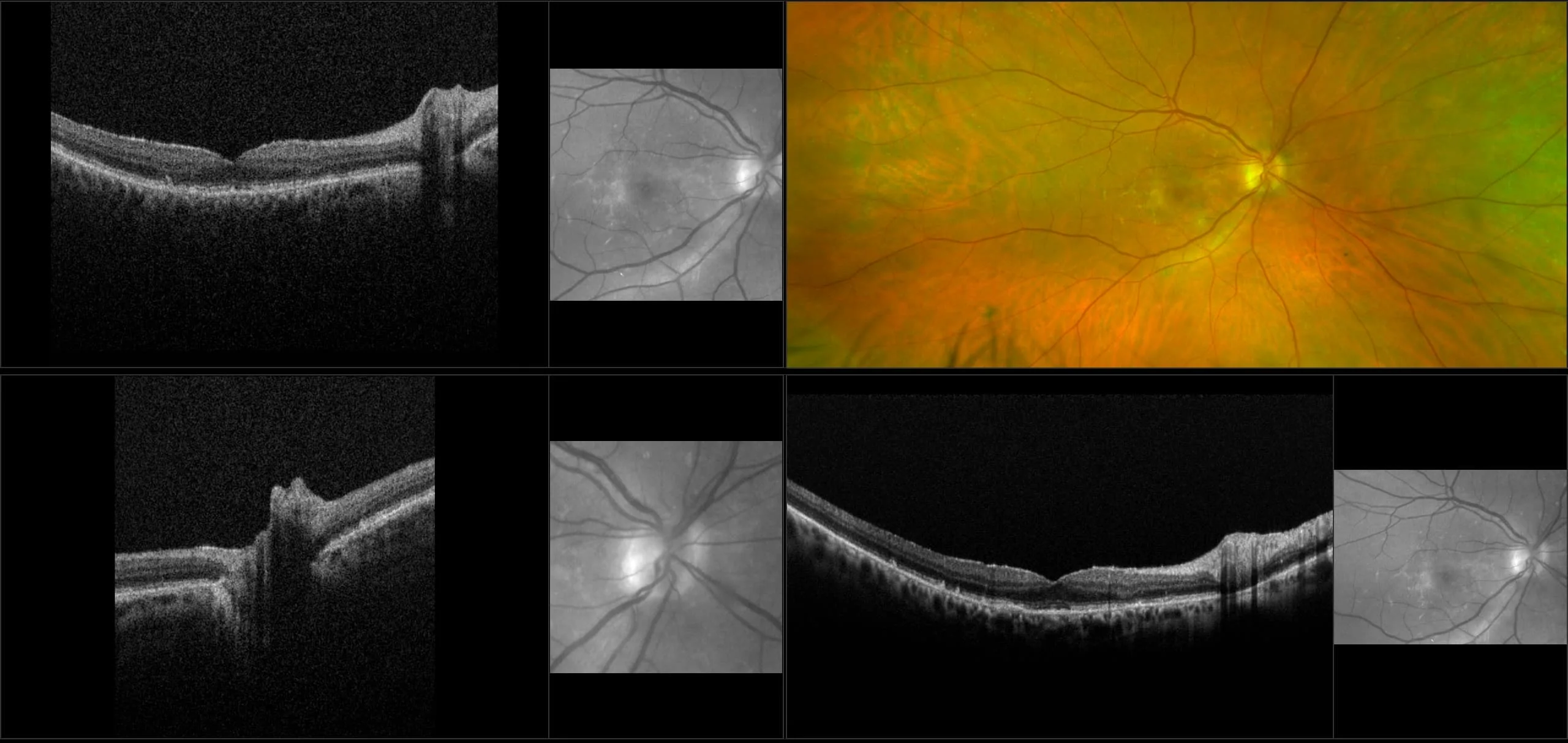
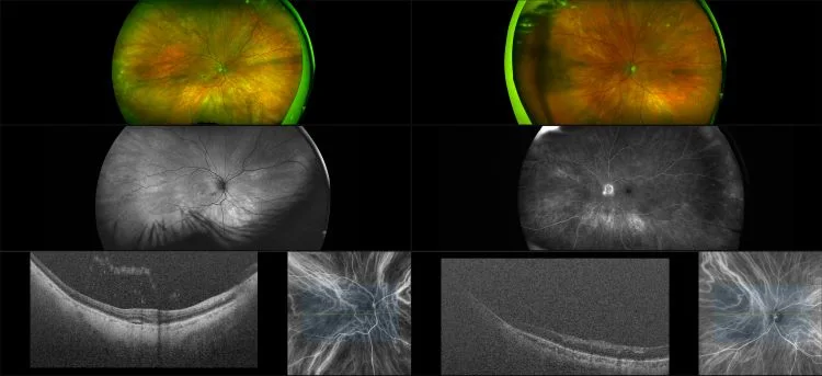
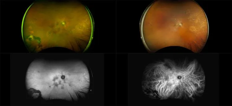
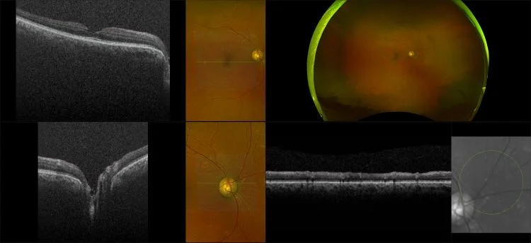
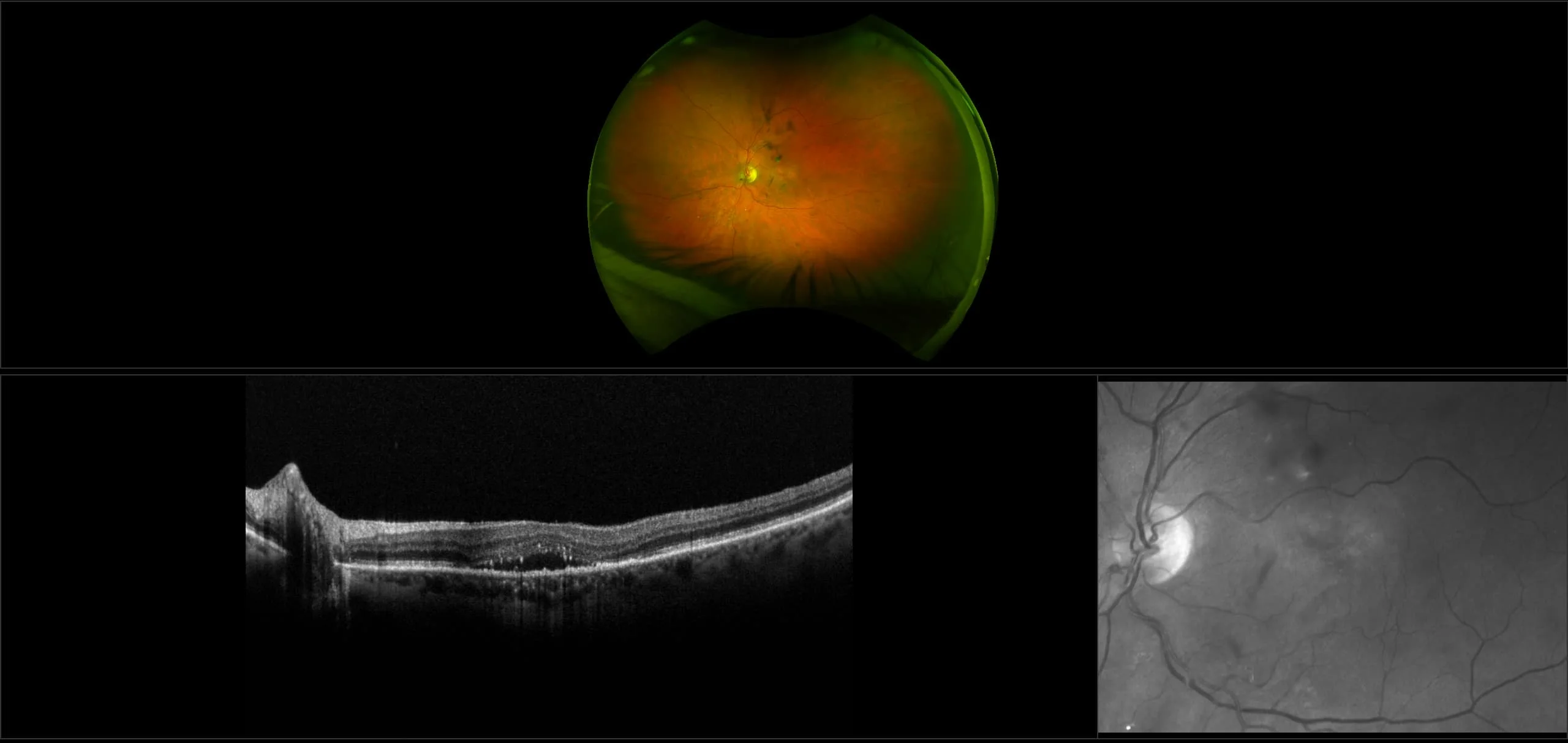
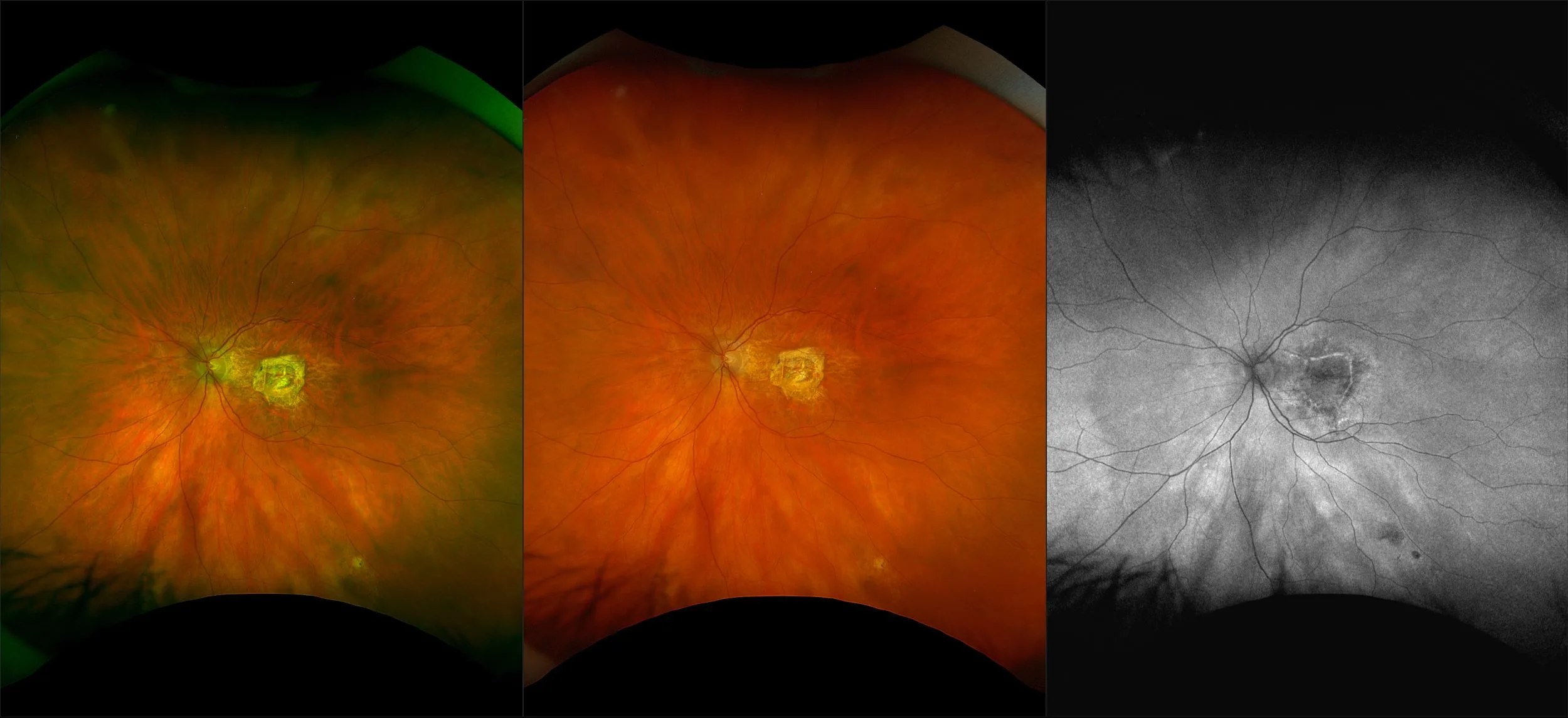
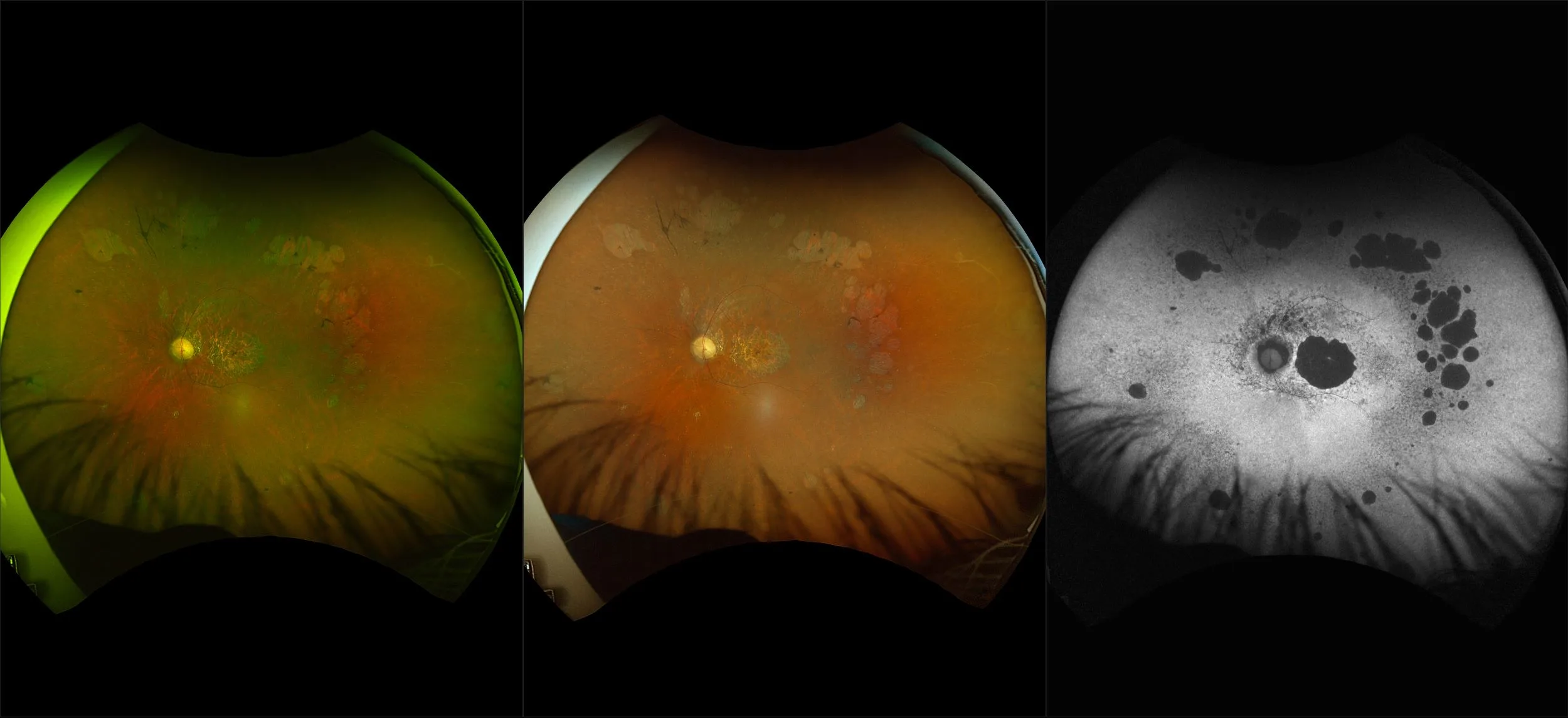
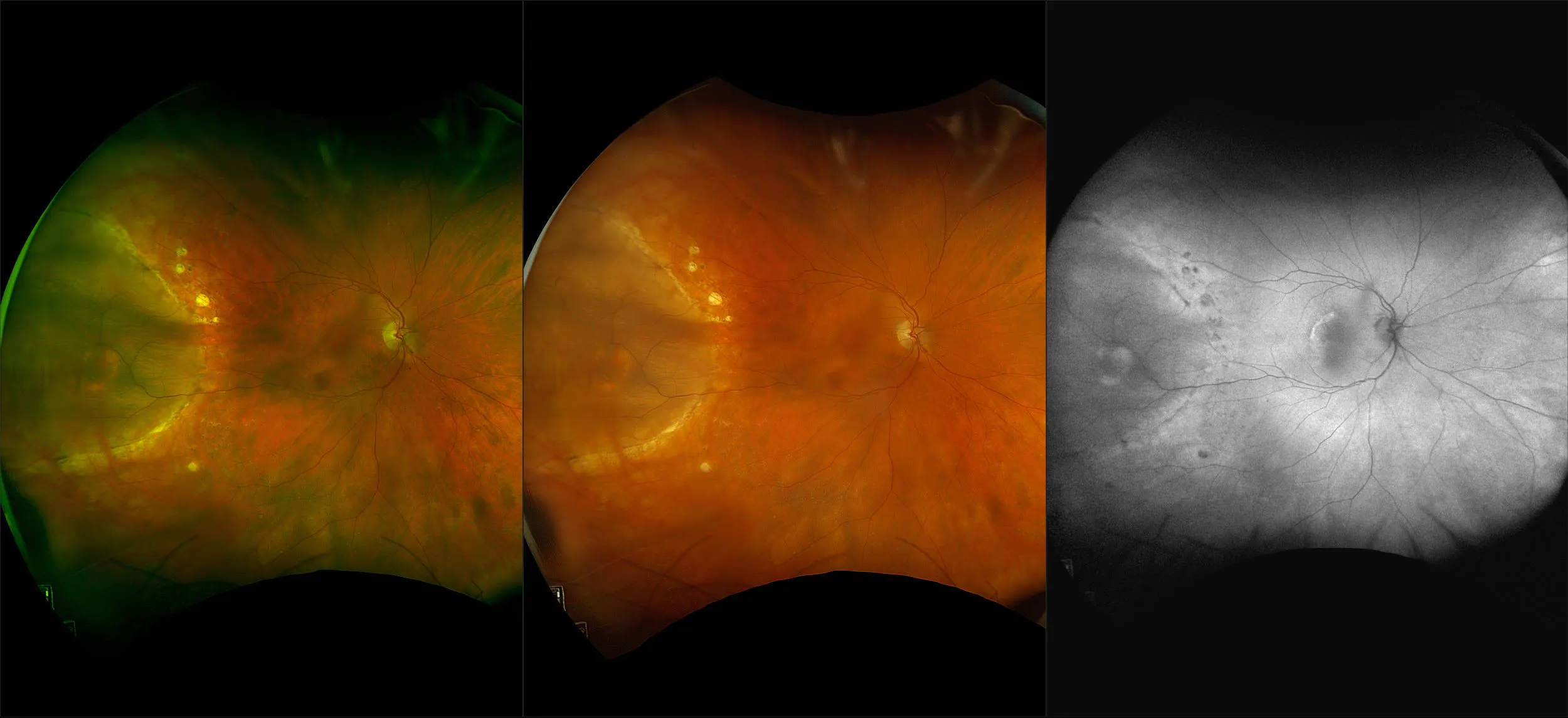
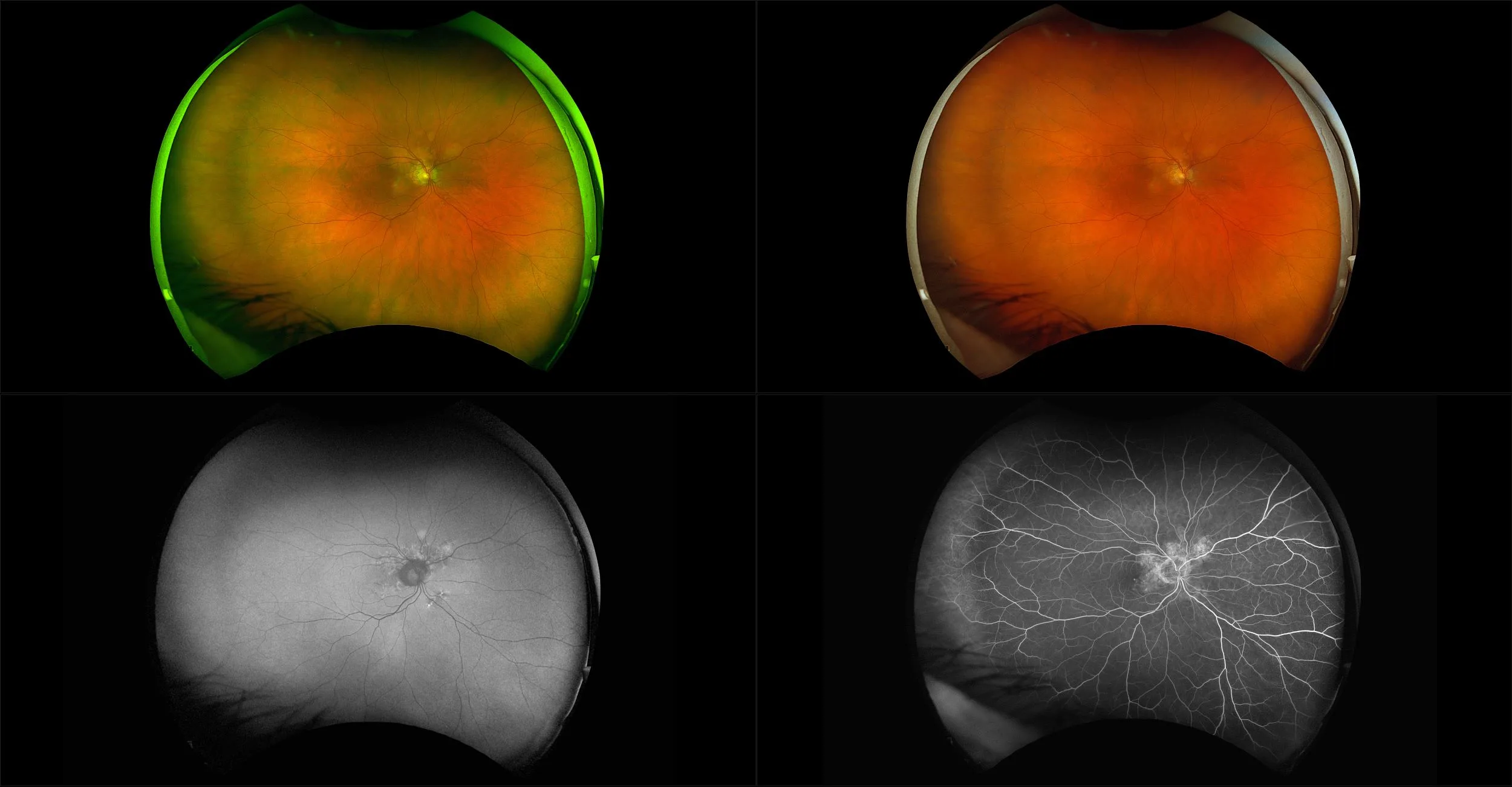
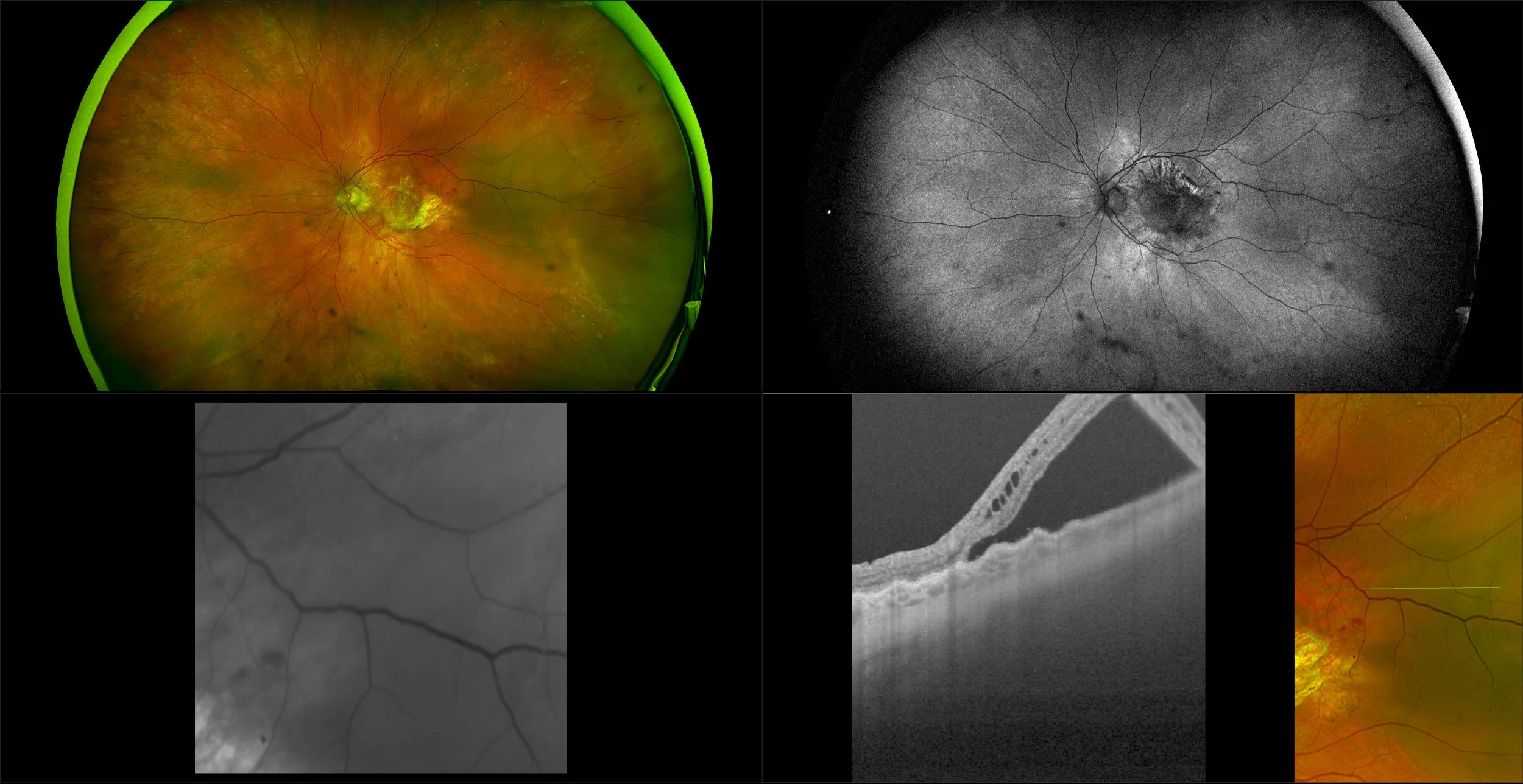
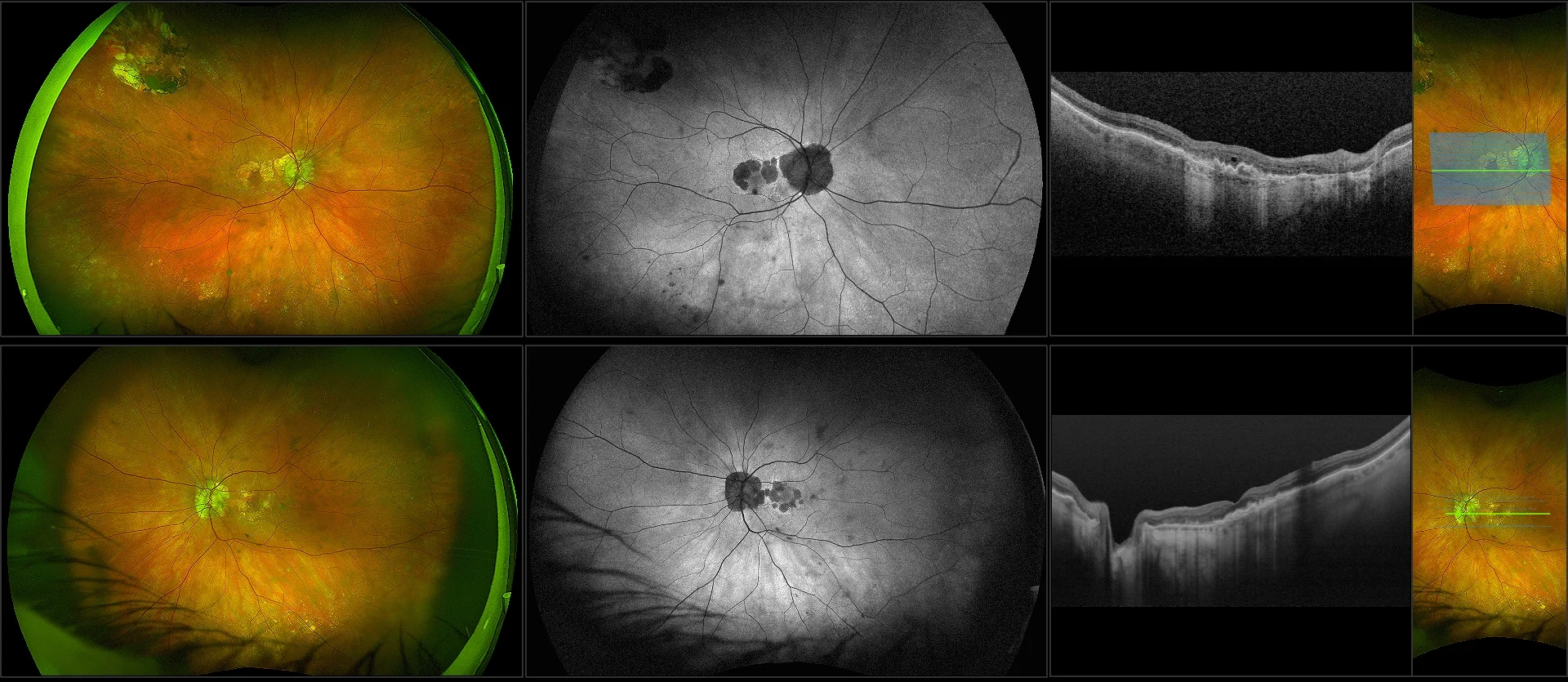
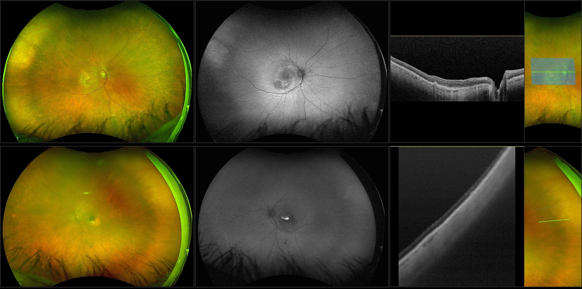
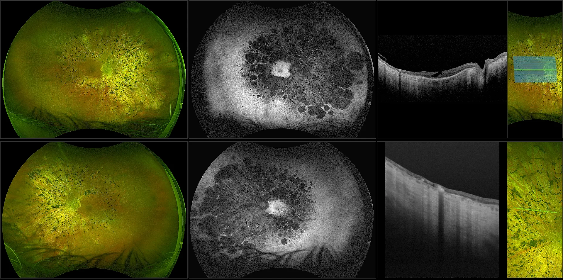
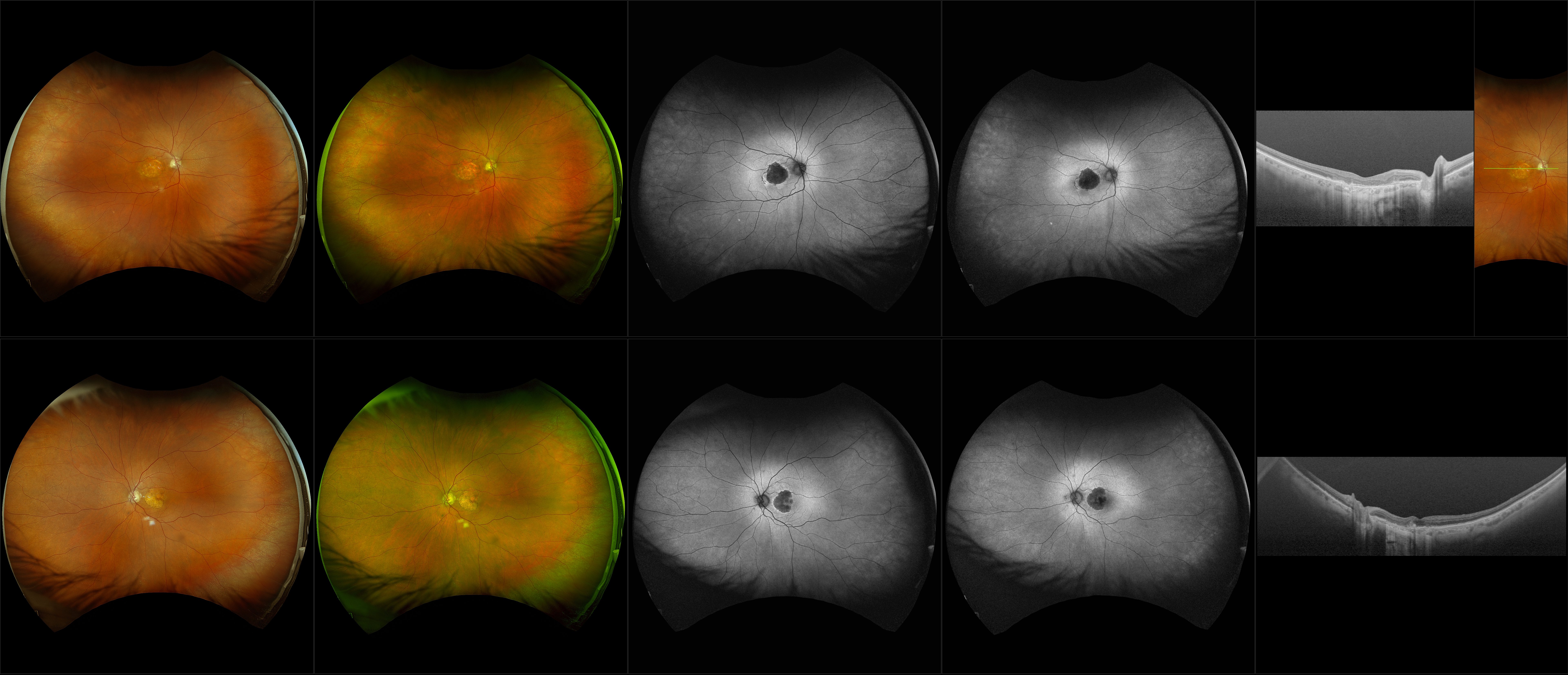
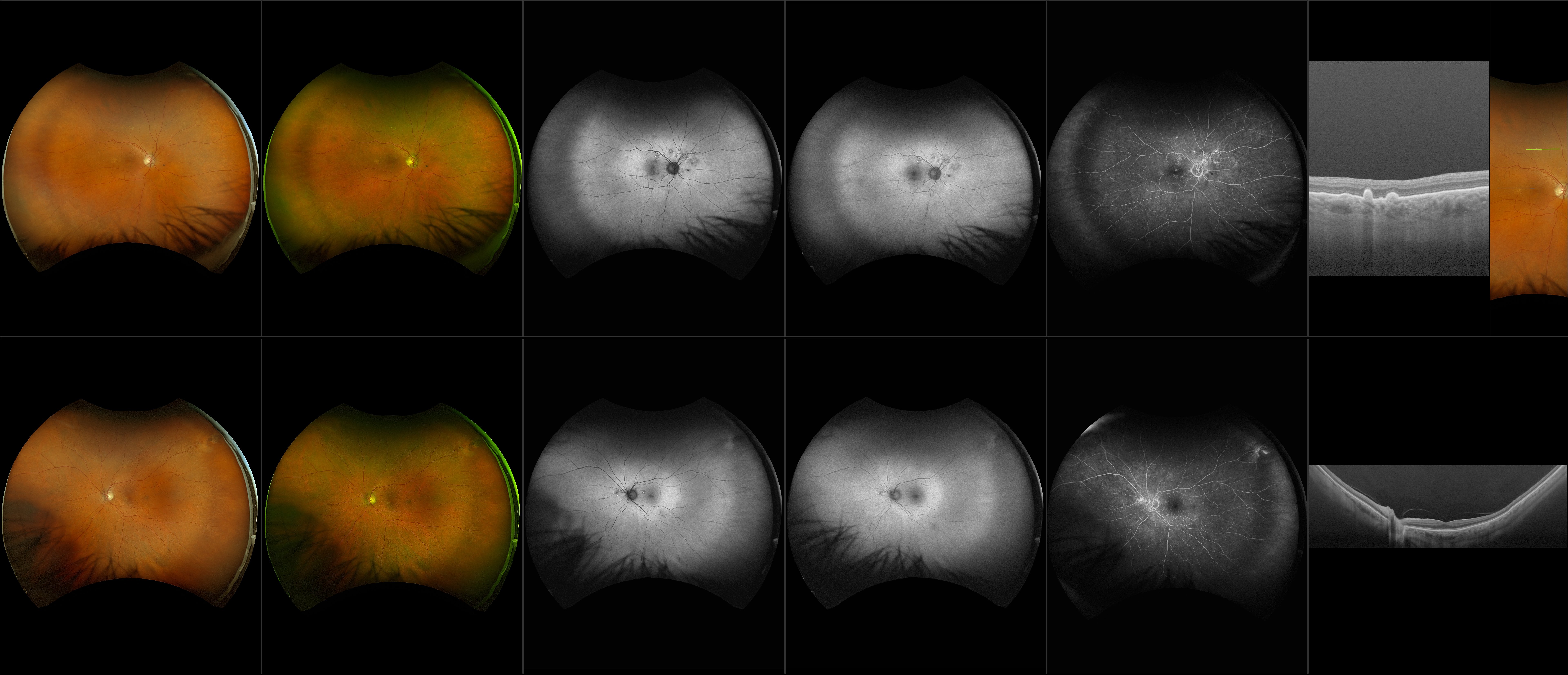
optomap Multimodal AMD Cases
Optos offers multimodal imaging with all ultra-widefield devices. Having both ultra-widefield and four images captured in less than one second has been shown to enhance pathology detection and disease management as well as improve practice and clinic flow. Ultra-widefield multimodal imaging is important across all access points of patient care - screening, detection, diagnosis, and treatment.
Related Cases
Other Diagnostic Tools