News & Press Releases
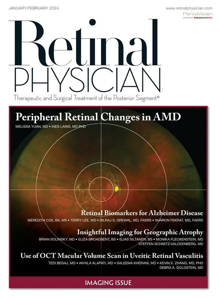


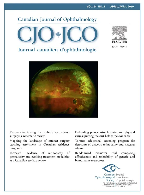


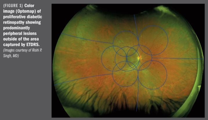
On the Cover: Boston grid superimposed on an UWF, optomap fundus image. From “Peripheral Retinal Changes in AMD,” page 12 (Retinal Physician).
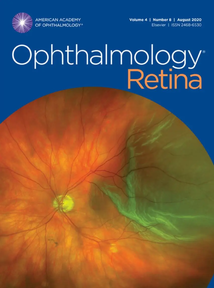
On the cover: “Healing Retinectomy Edge After Schisis-Detachment Repair” by Sarah Skiles, CRA and Jonathan Russell, MD (University of Iowa Hospitals and Clinics, Iowa City, IA). Equipment: Optos California.
Image compilation including optomap ultra-widefield retinal imaging.
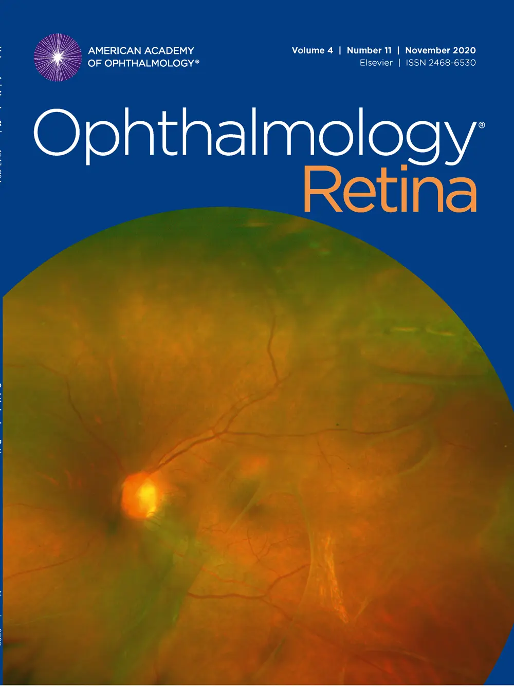
On the Cover: "Successfully Radiated Retinoblastoma after 30 Years" by David H. Abramson, MD, FACS, Jasmine H. Francis, MD, FACS, David K. Mock, COA, and Monica Adu (Memorial Sloan Kettering Cancer Center, New York, NY). Equipment: Optos (California).
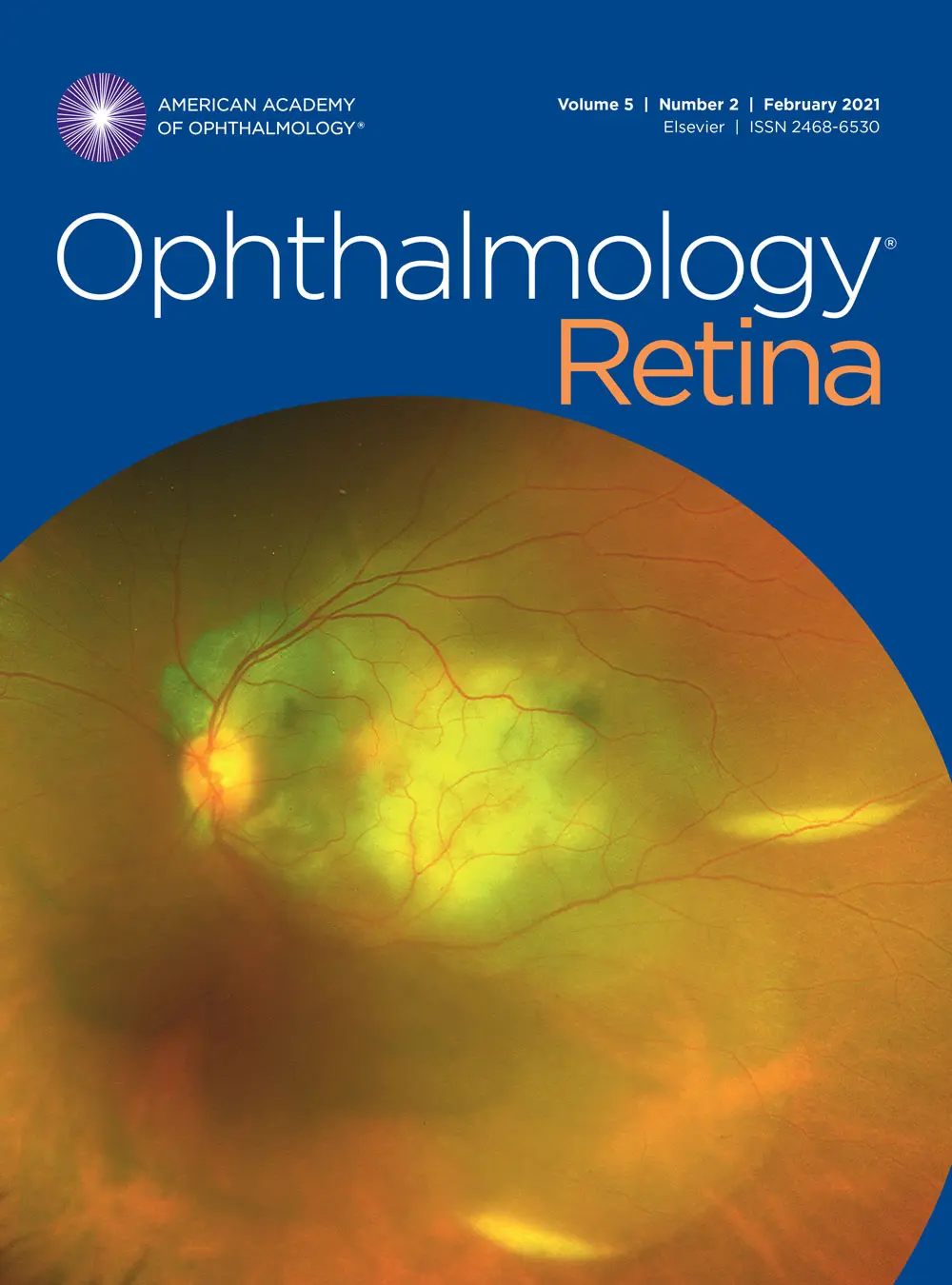
On the Cover: “What may lie beneath: A look at AMD with ICG angiography” by Dena Harris, CRA (Kellogg Eye Center University of Michigan, Michigan Medicine, Ann Arbor, MI) was the Second Place winner in the Indocyanine Green Angiography category at the Ophthalmic Photographers’ Society 2021 Scientific Exhibition (www.opsweb.org). Equipment used: OPTOS California.
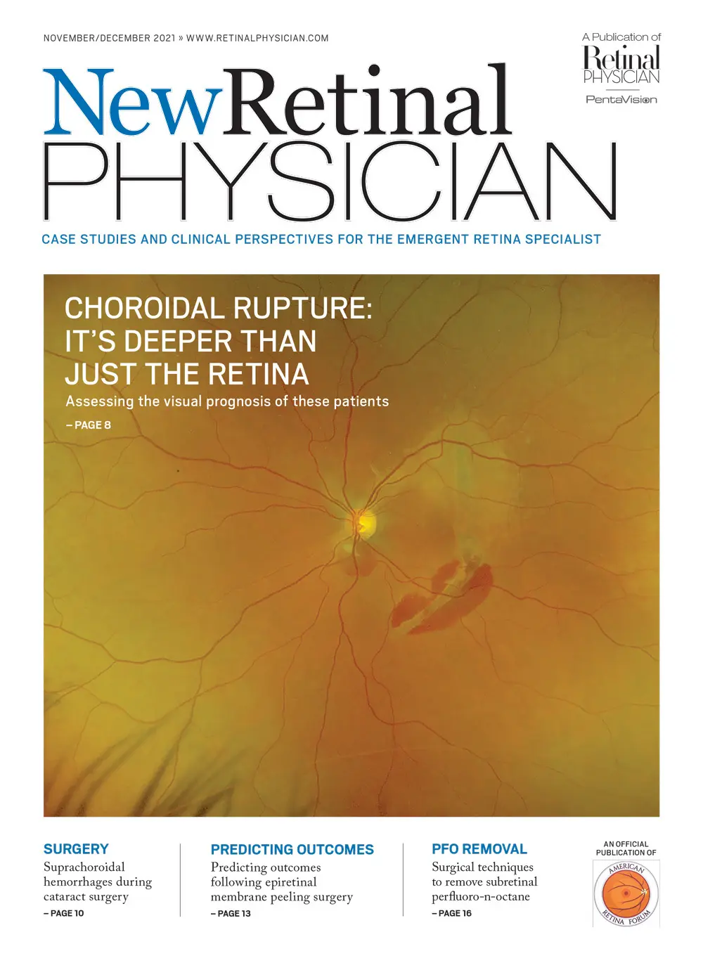
On the Cover: Choroidal Rupture. Hemang K Pandya, MD, Dallas Retina Center.
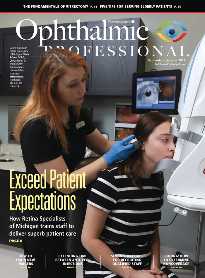
On the Cover: At Retina Specialists of Michigan, Olivia Rainey, OCT-C, COA, director of photography, demonstrates ultra-widefield imaging (California, Optos) with Brittany Rota, technician. Photo by David Sparks
On the Cover: Image courtesy of Optos.
On the cover: Choroidal metastasis from intrahepatic cholangiocarcinoma., y Saki Akou,CO, Photographer; Katsuhide Yamadera, CO, Photographer; Hisashi Fukuyama, MD, PhD, Clinician; Fumi Gomi, MD, Clinician (Hyogo College of Medicine, Hyogo, Japan).Equipment: Optos
Optos on the Cover: Drs. Salabati, Soares, and Hsu utilize optomap technology for their article “Loss to Follow-up After Anti-VEGF for AMD and DR”.
On the Cover: Color optomap image of Chronic birdshot chorioretinitis. Image courtesy of Pauline T. Merrill, MD.
On the Cover: Optos UWF image of proliferative DR with multiple pre-retinal hemorrhages, intraretinal dot and blot hemorrhages and superior/inferior scars from previous pan-retinal photocoagulation treatment. Images courtesy of Hannah Calvelli, O.D.
On the Cover: Image courtesy of Mitul Mehta, MD
On the Cover: Navigating the Retinal Periphery. Mohammad Rafieetary, OD
On the Cover: Double bubble posterior hypopyon in a patient with a presumed tubercularchoroidal granuloma”by Deeksha Katoch, M.S.; Vipin Rana, MS, FICO, MRCS (Ed);Atul Arora, MS; Arun Kapil, Chief Retinal Photographer (Advanced Eye Centre, PostGraduate Institute of Medical Education and Research, Chandigarh, India). EquipmentUsed: Optos California ultra-widefield imaging 200 DTx icg, Scotland, United Kingdom.
On the Cover: An optomap fundus photograph showing peripheral exudative hemorrhagic chorioretinopathy. From “Choroidal Melanoma or Peripheral Exudative Hemorrhagic Chorioretinopathy,”
On the Cover: “Vitreoretinopathy in a patient with Stickler syndrome. ”Veil-like vitreous condensations are apparent posteriorly along with patches of perivascular lattice. Nikita Gupta, MS; Atul Arora, MS; Ramandeep Singh, MS; Arun Kapil, Chief Retinal Photographer (Advanced Eye Centre, Post Graduate Institute of Medical Education and Research, Chandigarh, India). Equipment: Optos ultra-widefield imaging 200 Tx, V2 Vantage Pro software.
On the Cover: Fundus photograph of delayed-onset retinal detachment in the setting of postoperative Streptococcus intermedius endophthalmitis. From “Management of Concurrent and Delayed-Onset Retinal Detachment Associated With Endophthalmitis,” page 14.
On the Cover: Ultrawidefield fluorescein angiography (Optos) of a 47-year-old female with hemoglobin SC sickle cell disease showing sea-fan neovascularization in the peripheral retina of the left eye.
Volume 4, Number 8
On the cover: “Retinal Detachment” by James Gilman, CRA, FOPS (Moran Eye Center, Salt Lake City, UT) was Honorable Mention in the Color Fundus Ultra-Wide Angle category at the Ophthalmic Photographers’ Society 2019 Scientific Exhibition (www.opsweb.org). The patient is a 60 year-old white male with a history of macula off rhegmatogenous retinal detachment who later underwent pars plan vitrectomy, cryo, SF6. Equipment: Optos California.
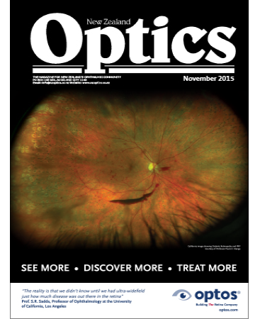
On The Cover: Monaco from Optos, ultra-widefield Image reveals moderate NPDR with peripheral
lesions and no DME. Image courtesy of Michael Singer MD.
On the Cover: optomap image showing Dry AMD. Elizabeth Steele, OD
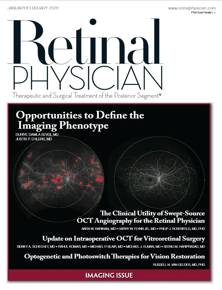
On the Cover: Opportunities to Define the Imaging Phenotype Duriye Damla Sevgi, MD and Justis P Ehlers, MD
On the Cover: Retinal Capillary Hemangioblastoma–"Double Disc Appearance" by Ram-andeep Singh, MS, Priya Bajgai, MS, Arun Kapil, Chief Retinal Photographer (AdvancedEye Centre, Post Graduate Institute of Medical Education and Research, Chandigarh,India). Equipment: Optos P200Tx
On the Cover: Single-capture, ultra-widefield optomap icg image of an eye with birdshot choroiditis. The white circle marks the locations of vortex vein ampullae in all four quadrants. The International Widefield Imaging Study Group definition of an UWF image is one that includes retinal anatomy anterior to the vortex vein ampullae, in all four quadrants. Image courtesy of Optos.
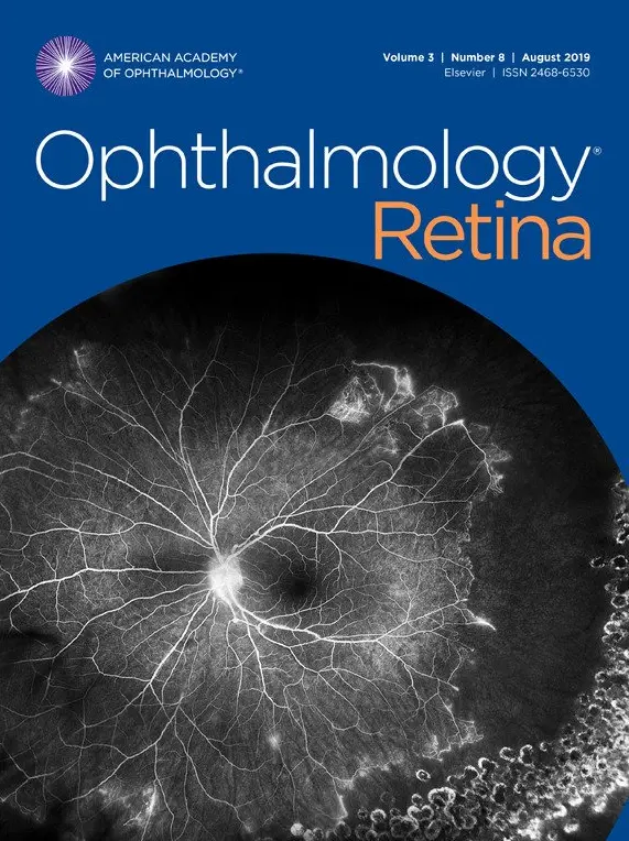
August 2019
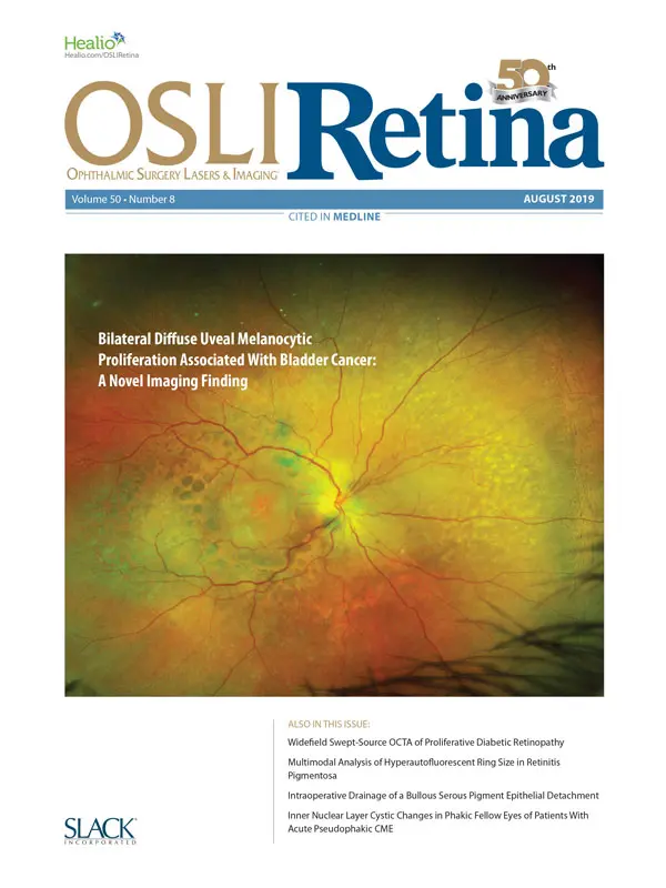
On the Cover: Bilateral Diffuse Uveal Melanocytic Proliferation Associated With Bladder Cancer: A Novel Imaging Finding. Roderick O'Day, MBBS; Kira Michalova, MD, FRANZCO; William G. Campbell, MRCP (UK), FRANZCO
On the Cover
Peripheral ischemia with I.R.V.A.N. (Idiopathic retinal vasculitis, aneu-rysms, and neuroretinitis) Five Optos California Ultra-wide images were montaged inPhotoshop so that all of the retina could be viewed in a single image, by Chris Barry (LionsEye Institute, Australia). Third Place winner Fluorescein Angiogram category at theOphthalmic Photographers’Society 2018 Scientific Exhibition (www.opsweb.org).
On the cover: Ultra-widefield image (Optos) shows diabetic retinopathy hemorrhages in the periphery. Image courtesy David Brown, MD.
ON THE COVER: A 14-year-old female with lesions suggestive of choroidal melanoma who underwent transvitreal fine-needle
aspiration biopsy. This image shows a small area of hemorrhage on the site of puncture, which resolved. See “Diagnostic Biopsy of
Intraocular Tumors”.
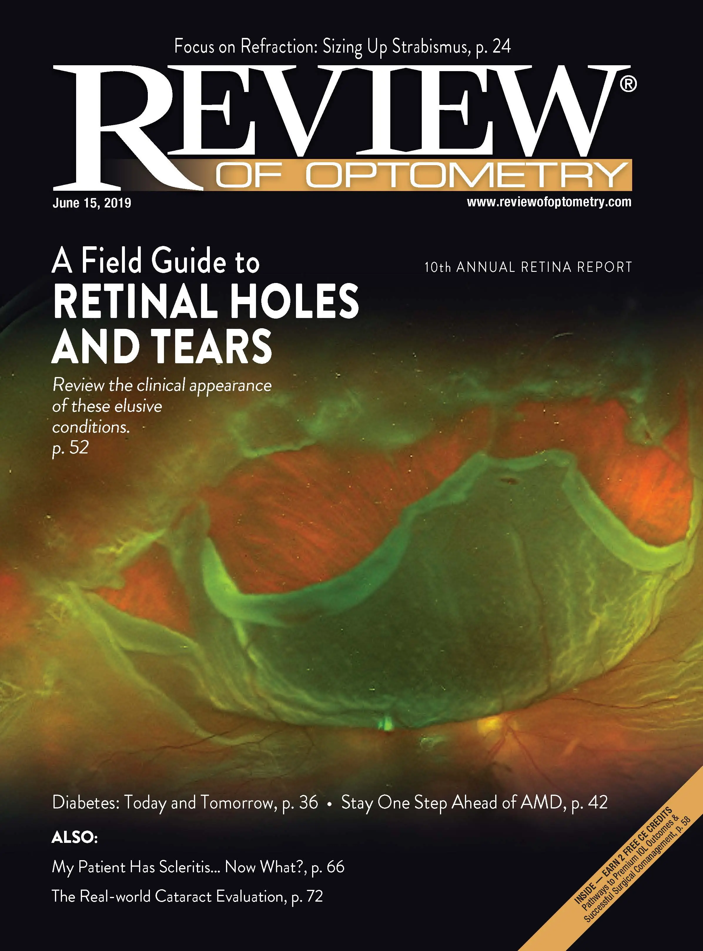
On the Cover: A Field Guide to Retinal Holes and Tears. Mohammad Rafieetary, OD and Stephen Huddleston, MD
Cover Image: Fundus Autofluorescence in ABCA4-gene associated Stargardt Disease. Submitted by Denise A Krolnik (Photographer), Kathleen A Regan MD, and Kimberly E Stepien MD. (University of Wisconsin-Madison, Department of Ophthalmology and VisualSciences). Equipment: Optos 200Tx with ResMax
Cover Image: Emulsified silicone oil implant forming a horizontal level in the posterior segment, several years after implantation with retinal detachment repair. From a team at Dalhousie University School of Ophthalmology & Visual Sciences.
Cover Image: Provided by Dean Eliott, MD, a member of the Retina Today Editorial Advisory Board, and Kareem Moussa, MD. Dr. Eliott is the Stelios Evangelos Gragoudas Professor of Ophthalmology at Harvard Medical School. Dr. Eliott is also the Director of the Retina Service and the vitreoretinal fellowship at Massachusetts Eye and Ear. Dr. Moussa is a second-year vitreoretinal fellow at Massachusetts Eye and Ear. The photographers for this case were John Hensel, OCT-C; and Roy Salvador, COA, who captured the images during two sessions at Massachusetts Eye and Ear.
Cover Image: “Giant Retinal Tear” by James Gilman, CRA, FOPS (Project Administrator for the Moran Eye Center) was the Third Place winner in the Ultra-widefield Imaging at the Ophthalmic Photographers’ Society 2017 Scientific Exhibition (www.opsweb.org<http://www.opsweb.org>). Equipment: Optos California.
On the cover: Surgical Management of Proliferative Diabetic Retinopathy
Considerations and treatment approaches. MICHAEL J. VENINCASA, BS • NANDINI VENKATESWARAN, MD • JAYANTH SRIDHAR, MD
Cover Image: “Asteroid Hyalosis” by James Gilman, CRA, FOPS (Project Administrator for the Moran Eye Center) was the First Place winner in the Ultra-widefield Imaging at the Ophthalmic Photographers’ Society 2017 Scientific Exhibition (www.opsweb.org). Equipment: Optos California.
On the Cover: Saturday night retinopathy after intranasal heroin. Top: (Left) Widefield fundus photo of affected right eye displaying diffuse retinal whitening without cherry red spot. (Right) Fluorescein angiogram displaying patchy choroidal filling and poor macular filling. Bottom: Brain MRI FLAIR imaging displaying diffusion restriction of optic nerve (arrow in left photo) and bilateral hippocampal diffusion restriction and swelling (arrows in right photo). (Photos courtesy of Huy V. Nguyen, MD, Victoria S. North, MD, Patrick Oellers, MD, and Deeba Husain, MD.)
On the Cover: Ultra-widefield fundus photograph of a retinal detachment due to macular hole in a high myopic eye, from “Macular Buckling Technique in High Myopia.”
Image courtesy of Micol Alkabes, MD and Carols Mateo, MD
On the Cover: Optos preoperative photo of the right eye of a 35-year-old woman with type 1 diabetes who experienced progressively decreased visual acuity to hand motions. A large frond of fibrovascular membranes is seen superotemporally, causing a tractional retinal detachment in that area. A peripheral tractional and rhegmatogenous retinal detachment is seen from 12 o'clock to 8 o'clock. The patient underwent a bevacizumab injection 3 days before vitrectomy 1 month post 27-gauge vitrectomy with probe-only lift, shave dissection techniques, and 1,000-centistoke silicone oil tamponade. The retina is fully attached, and the best corrected visual acuity is 20/70.
Image Courtesy of MARÍA H. BERROCAL, MD • LUIS A. ACABA, BA
On the Cover: Bullous serous retinal detachment from uveitis.
Image courtesy of Tara A. McCannel MD, PHD
Cover Image: Ultra-widefield fundus photograph (optomap) of postoperative day 1 showing subretinal air with complete displacement of the subretinal hemorrhage inferiorly (sitting position), and partial gas fill in the vitreous cavity, preventing the subretinal air from migrating superiorly to keep it more posterior and allow complete displacement.
From Sharma S, et al, “Pneumatic Displacement of Submacular Hemorrhage with Subretinal Air and Tissue Plasminogen Activator: Initial United States Experience” on page 180 in this issue.
Cover Image: "Argus Retinal Prosthesis" by Robert Prusak, CRA (Kellogg Eye Center, Ann Arbor, MI) was the Second Place winner in the Instrumentation category at the OPS 2016 Scientific Exhibition. The prosthesis is implanted in a patient with retinitis pigmentosa.
Imaged on a 200Tx from Optos.
Cover Image: An optomap ultra-widefield color fundus image of the right eye showing two pigmented choroidal masses at diagnosis with orange pigment overlying both lesions.
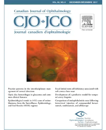
Image provided by: Padron-Perez N, et al., "Bilateral multiple iridociliary cysts in diffuse uveal melanocytic proliferation", Canadian Journal of Ophthalmology, Volume 52, Number 6.
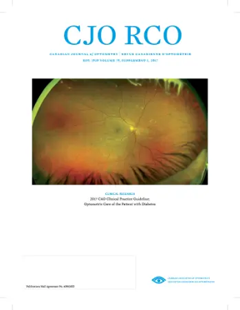
Cover Image: 2017 CAO Clinical Practice Guideline: Optometric Care of the Patient with Diabetes (image supplied by Optos, Inc and taken by Professor Paolo Stanga.)
Cover Image: Uveitis with Progressive Subretinal Fibrosis in a Young Child e Starry Sky Appearance on the Retina. A 10-year-old female child with delayed development and taking oral anti-epileptics presented with decreased vision in both eyes, right worse than left. On examination, there were multiple round lesions over the posterior pole of the fundus with subretinal fibrosis. Image captured using Optos 200Tx.
Imaged By:
Arun Kapil, Advanced Eye Centre, Post Graduate Institute of Medical Education and Research, Chandigarh, India
Cover Image: “Retinal Detachment” by Sarah M Armstrong, CRA, OCT-C, FOPS (University of North Carolina Kittner Eye Center, Chapel Hill, NC) received Honorable Mention in the Ultra-Widefield Imaging category at the Ophthalmic Photographers’ Society 2016 Scientific Exhibit (www.opsweb.org).
This image was obtained with the Optos California (Optos plc, Dunfermline, Scotland, UK). During this initial visit, patient was seen by Michelle Go, MD; Kevin Gertsch, MD; and Ali Torab Parhiz, MD.
Cover Image: “Fovea-sparing Choroideremia” by Tim Steffens (University of Michigan Kellogg Eye Center) was the Second place winner in Cross Categories at the Ophthalmic Photographers’ Society 2016 Scientific Exhibition (www.opsweb.org).
Fundus, optical coherence tomography (OCT), and autofluorescence images of a 38-year-old man with choroideremia-like phenotype (CHM gene mutations negative). The fundus shows extreme loss of choriocapillaris and retinal pigment epithelium (RPE) leaving intact islands of retina. The OCT shows loss of RPE by the signal penetrating through into the choroid.
Cover Image: Color fundus photograph of bird-shot retinochoroiditis in a 62-year-old woman Courtesy Vicky Wolzen, CRA, COA, Dean McGee Eye Institute.
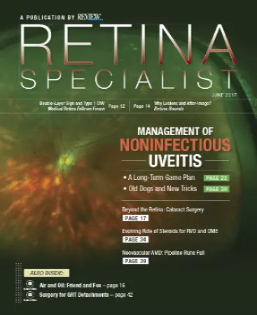
Content provided for article, A Long-term Game Plan for Noninfectious Uveitis, article written by, Sam S. Dahr, MD
Cover Image: Ultra-widefield color imaging showing angioid streaks around the optic disc in a 47-year-old man with pseudoxanthoma elasticum. Comet lesions are visible in the retinal periphery. Adapted from Marchese A et al, “Ultra-widefield imaging in patients with angioid streaks secondary to pseudoxanthoma elasticum” on page 137 in this issue. Color ultra-widefield fundus photo captured by optomap California fundus camera (Optos PLC, Dunfermline, UK).
Photographers: Adriano Carnevali, MD, and Lea Querques, MD, Department of Ophthalmology, IRCCS Ospedale San Raffaele, University Vita-Salute San Raffaele, Milan, Italy.
Cover Image: Gaucher’s disease. Baseline photograph OD taken of a 15-year-old male with a history of strabismus, bilateral 6th nerve palsy, and Gaucher's disease. Macula and vessel findings were observed to be normal. However, ultra-widefield imaging revealed numerous white deposits of varying size found in all four quadrants of the midperipheral retina bilaterally. The image was captured using Optos P200Tx.
Imaged by: Ryan Imperio, CRA, OCT-C
Cover Image: (A,B) Ultra-widefield color fundus and autofluorescence photos of the left eye of a 69-year old woman with a history of ROP showing typical pigmentary changes and patches of lattice degneration. (C-E) There is an area of tractional retinoschisis in the temporal macula associated with dense hyaloidal organization. (F,G) Ultra-widefield color fundus photo of the right eye of a 65-year old patient with history of ROP. Notice the characterisic straightening of the vessels and peripheral pigmentary changes in the temporal macula. (H) There is tractional retininoschisis along the superior temporal vascular arcade. (I,J) Preservation of inner retinal layers after laser photocoagulation with associated choroidal excavation (arrows).
Paper and images by: Aristomenis Thanos, MD, et al.
Cover Image: Clinical features of DR may include hemorrhages, hard exudates, cotton wool spots, and/or neovascularization.
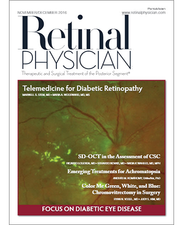
Paper and image by: Maxwell S. Stem, MD and Maria A. Woodward, MD, MS.
Cover Image: “Double Bubble Trouble.” Intravitreal live dual. Cysticercus cysts with scolex protruding out of the cyst along with total retinal detachment.
Submitted by: Dr Ravi Bypareddy and Rohan Chawla; Photographer: Kabiruddin Molla, Bsc (Optometry) and Dr. Karthikeyan Mahalingam. Affiliation: Dr Rajendra Prasad Centre for Ophthalmology, All India Institute of Medical Sciences, New Delhi, India 110029. Equipment: Optos 200Tx.
Cover Image: Optos UWF image of a patient with newly diagnosed type 2 diabetes. This image reveals significant pathology, including ischemia and neovascularization outside the traditional imaging area. This patient was treated with targeted panretinal photocoagulation using the UWF FA as a guide for lasering the areas of nonperfusion.
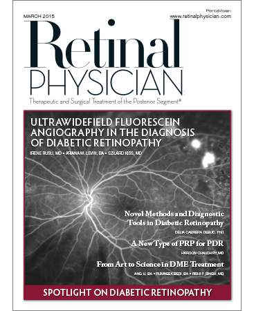
Paper and image by: Irene Rusu, MD et al.
Cover Image: Image shows a patient with an inferior retinal detachment, shallow submacular fluid associated with hard exudates and subretinal fibrosis. Creamy subretinal infiltrates along the superotemporal arcade also visible. Optical coherence tomography with a cut through the superotemporal arcade showed dialated choroidal vasculature, multiple pigment epithelial detachments and hyper reflective subretinal infiltration.
Paper and images by: Duncan Berry, MD et al.
Cover Image: optomap image courtesy of Optos
The Daytona is significantly smaller than previous devices. The technology also captures FAF images. In addition, the Daytona and California (shown) image 82% or 200 degrees of the retina in a single capture.
Cover image: optomap image courtesy of Optos
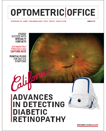
Cover Image: California image showing Diabetic Retinopathy and PRP.
Imaged by: Professor Paulo E. Stanga
Cover Image: shows an 8mm x 6mm mottled melanotic choroidal nevus with no appreciable thickening in the superior mid-periphery, with overlaying drusen and intra-retinal hemorrhaging. There are also intra-retinal hemorrhages extending into the superior periphery. The lesion has no evidence of orange pigment or subretinal fluid.
Paper and images by: Sara Weidmeyer, OD et al.
Cover Image: Primarily peripheral lesions in a patient with moderately severe nonproliferative diabetic retinopathy. This patient has high risk of progression and proliferative disease.
Paper and images by: A. Paul Chous, MA, OD.
On the Evolution of Retinal Laser Photocoagulation.
Cover image: optomap image courtesy of Optos
On the cover: “Giant Retinal Tear” by Govindarajan Jayaraman (Senior Ophthalmic Photographer, Seri Research Clinic, Singapore Eye Research Institute, Singapore) was the Honorable Mention winner in the Ultra-Widefield Imaging category at the Ophthalmic Photographers’ Society 2021 Scientific Exhibition. Equipment: Optos California.
Press releases, select articles, peer-reviewed papers, and key clinical findings. Listed chronologicially by year.
2026
2025
Optos unveils Silverstone RGB, setting a new standard for retinal imaging technology. The only device combining 200° single shot true-to-life color imaging with advanced swept-source OCT
Optos, a leading provider of ultra-widefield retinal imaging technology, today announced that it has achieved both Stage 2 ISO 27001:2022 certification and Authority to Operate (ATO) from the U.S. Department of Defense (DoD)
2024
2023
Optos, Plc, the leading retinal imaging company, today announces the advances of Ultra-widefield Multimodal Retinal Imaging, a central focus of Optos’ satellite symposia at EURETINA 2023.
Optos, Plc, the leading retinal imaging company, today announces, it is expanding the optomap® ultra-widefield (UWF™) retinal imaging modalities available with the California FA device to further assist Eyecare Professionals in disease management and treatment planning.
2022
Optos, a Fife-based company, has reported significant growth with record group revenue of $254m in the year to March 22 and more than 22,000 devices installed worldwide.
2020
Optos, a Fife based company and a world-leader in the design and manufacturing of advanced retinal imaging technology, has been awarded NHS funding to accelerate the implementation and validation of Artificial Intelligence (AI) to support the early detection of Diabetes related eye-disease into NHS Diabetic Eye Screening practices.
2019
A review of the International Widefield Imaging Study Group recommendations for a terminology to describe image captures from various modalities.
Optos plc, the leading medical retinal imaging company, part of Nikon Corporation, is pleased to announce the launch of Silverstone at the American Academy of Ophthalmology, San Francisco, CA.
Given the well-documented relationship between posterior pole nonperfusion and DR progression, it is logical that greater peripheral nonperfusion on ultra-widefield fluorescein angiography is also associated with higher likelihood of PDR.

Dunfermline-based Optos, a leading provider of retinal imaging devices, kept its crown at this year’s Optician Awards in Birmingham (30th March), claiming the title of Optical Supplier of the Year for the second time in a row.
2018

Ultra-widefield fluorescein angiography revealed distinct fundus-wide patterns of vascular damage, which were progressive in nature in eyes treated with iodine-125 brachytherapy for uveal melanoma and correlated with signs of progressive vascular injury. This grading scheme may have prognostic value to predict the progression of radiation retinopathy and to prognosticate visual outcomes in patients undergoing brachytherapy.
optomap UWF technology from Optos, is the only technology that can image beyond the vortex ampullae (up to 200° or 82% of the retina) in a single capture. optomap is available on all of our devices in all modalities and with Monaco, it can be paired with integrated spectral-domain OCT.
There's new eye equipment that makes finding potential vision problems easier and more. The first advantage with this device is that it eliminates the need for your eyes to be dilated when doctors look for things like glaucoma and cataracts and it's also helping doctors detect serious health issues.
Optos Plc, a subsidiary of Nikon Corporation, Japan, and Amydis, Inc. today announced a clinical alliance focused on the development of an eye test by Amydis to detect Alzheimer’s disease. Amydis has developed a pipeline of compounds to detect amyloid proteins in the retina to be visualized with Optos’ market-leading optomap ultra-widefield retinal imaging devices to diagnose Alzheimer’s Disease in patients.

Researchers from Queen’s University Belfast have shown for the first time that the eye could be a surrogate for brain degeneration like Alzheimer’s disease (AD).
These research results have recently been published in the Journal of Ophthalmic Research and is the first clinical study showing a potential for peripheral retinal imaging to be used in monitoring AD and potentially other neurodegenerative diseases.
The team, led by Dr Imre Lengyel, Senior Lecturer and Researcher at the School of Medicine Dentistry and Biomedical Sciences at Queen’s University have found that by examining the eye we might be able to reflect on what might be taking place in the brain.
The work was carried out alongside health professionals and care providers for AD patients and explored whether there are manifestations of AD in the eye.
Based on laboratory observations the team hypothesized that changes in the peripheral retina could be important to explore the association between the eye and the brain.
Using ultra-widefield imaging technology developed by Optos Plc, the team found that there are indeed several changes that seem to be, especially in the peripheral retina, associated with this debilitating condition.
Ultra-widefield angiography has been successfully used without sedation for evaluation of children with various pediatric retinal diseases such as Coats disease, familial exudative vitreoretinopathy, and retinopathy of prematurity.
Today, Monday 14th May 2018, Optos are joining BBGR (Nikon Lenswear France)* for their annual Vision Van – the ‘Tour de France’ to highlight the importance of regular eye tests to maintain good vision. The Vision Van is an optometry practice on wheels fully equipped with an Optos Daytona plus and full eye testing equipment. The van will provide free eye tests for local communities in six towns and cities across France to educate visitors the importance of regular, thorough eye health checks.
Optos plc, the leading medical retinal imaging company, part of Nikon Corporation, is pleased to announce that Monaco, the first ultra-widefield (UWF) imaging device combined with OCT, was launched today in the United States.

As a world leading manufacturer of retinal imaging technology, Optos was crowned Optical Supplier of the Year at the Optician Awards 2018 in Birmingham on Saturday 14th April. The awards are recognised as the benchmark of excellence in the UK optical community and showcase the best in achievement, ability and performance.
Results suggest an association between Dark Adaptation and the presence of peripheral reticular pigmentary changes, as well as the presence of a peripheral mottled decreased FAF pattern. This provides new insights on the clinical significance of peripheral changes in AMD, and their contribution to impairments on Dark Adaptation.
UWF retinal imaging revealed a significant association between AD and peripheral hard drusen formation and changes to the vasculature beyond the posterior pole, at BL and
after clinical progression over 2 years, suggesting that monitoring pathological changes in the peripheral retina might become a valuable tool in AD monitoring.
After being among the finalists for ‘Product of the Year’ for Daytona plus at the 2018 AOP Awards, we are pleased to announce that Optos has been shortlisted as ‘2018 Optical Supplier of the Year’ at the upcoming Optician Awards, “the benchmark of excellence in the Optical community” (theopticianawards.co.uk).
This year’s awards are even more exciting for Optos as we learn that a range of our UK customers have also made the finals across several categories.
After conversion to ranibizumab (from bevacizumab) in eyes with persistent DME refractory to bevacizumab, significant functional and anatomic improvements were noted. Qualitative analysis of ultra-widefield fluorescein angiography images was performed to identify retinal vascular pathologic features, specifically by observing the presence of vascular leakage, neovascularization, macular ischemia, and peripheral ischemia. The prevalence of macular ischemia was 33.3% at baseline and remained stable at study completion. Finally, the proportion of peripheral ischemia observed in participants increased from 66.7% at baseline to 70.3% at month 12, although the incidence of any leakage remained stable at month 12.
Prospective, observational study including patients with sickle cell disease. All patients underwent dilated fundus examination by a fellowship-trained retina specialist, as well as UWF fundus photography (FF) and fluorescein angiography (FA). Sickle retinopathy stage and treatment recommendation per eye were determined after clinical examination, UWF-FF, and UWF-FA, respectively, and differences in retinopathy stage and treatment recommendation were compared.
Conclusion: Ultra-widefield imaging detects a higher stage of sickle cell retinopathy compared with clinical examination alone, but these differences may not be clinically significant.
The patient presented with 20 years of exposure to HCQ, at a daily dose of 5.2mg/kg of actual body weight, and manifested a pericentral-only phenotype of HCQ toxicity, as demonstrated with detailed structural and functional testing.
2017
optomap examinations are a convenient and feasible method to determine fundus pathological changes in myopic patients, especially for patients who can not endure pupil dilation. Compared to the Goldmann three-mirror contact lens examination, the detection rate with optomap was 91.73%% for retinal lesions, while the detection rate of mydriatic slit-lamp lens exams was 81.20%.
In a primary health care clinic, 632 healthy subjects underwent UWF imaging during annual health screening. optomap images detected peripheral retinal pathology in 18.4% that was not visible on corresponding single-field (45°) images. UWF imaging is feasible in a primary care setting, allows improved visualization of peripheral retinal pathology, and may therefore be useful for telemedicine screening.
Wide-field retinal imaging revealed the presence of peripheral pigmented retinal lesions resembling CHRPE lesions in a subset of patients with genetically confirmed Stargardt disease. Presence of these lesions may be associated with severe phenotypes of the disease.
The present study (1,024 subjects) has shown the effectiveness of DR screening using UWF fundus camera detecting 9.27% of patients with DR. It has also shown the effectiveness of trained nursing personnel taking fundus pictures. First, UWF imaging has a fast learning curve and it is quick to take the picture. Second, widefield view is greater than the 7-field photography enabling more DR lesions to be detected. And third, images can be manipulated in terms of red-free, infra-red and image zoom. Hence, the present telemedicine model has demonstrated that in a multi-specialty private hospital, DR screening can be effective using UWF fundus camera.
Peripheral vascular leakage (PVL) is a possible independent driver of treatment augmentation. Patients with PVL were four-times more likely to have treatment augmented after adjusting for baseline presence of CME, diffuse vascular leakage and clinical grading of uveitis activity. This suggests that PVL may have influenced the decision to augment therapy in patients.
UWFFA identifies retinal vascular pathology in intermediate uveitis not present on clinical examination. Diffuse retinal vascular leakage was associated with worse visual acuity when compared to peripheral and no leakage patterns.
Grading of UWF imaging has high reproducibility in evaluating vertical cup-to-disc ratio and agreement with stereoscopic optic disc imaging and may be suitable for glaucoma diagnosis in situations where colour digital stereoscopy is not available.
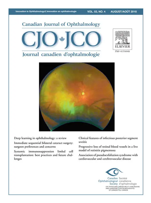
Peripheral retinal changes were observed peripheral to the equator in more than 40% of analyzed eyes in patients with and without AMD. Drusen, reticular pigmentary change and paving stone degeneration occur significantly more frequently in eyes with AMD compared to healthy eyes.
This year is the 25th anniversary for Optos. With such a significant milestone, it is customary to reflect on the journey. It all began when Douglas Anderson, our Founder set out to commercialize a patient-friendly product that encompassed a digital widefield image of the retina in a single capture. His young son, Leif was the impetus behind this mission. Whilst he had been receiving regular eye exams, the doctor was unable to examine Leif’s entire retina and he became blind in one eye when a peripheral retinal detachment went undetected. From that point on, the mission of saving sight and saving lives was engrained in the fabric of our culture and has become the cornerstone of our vision to be The Retina Company.
Specific AMD features in UWF images were graded. Standardised grids were used to record the spatial location of AMD features, including RPD. We found no evidence to support an association between coronary artery disease and RPD. RPD was strongly associated with intermediate AMD features.
Demonstrated that Coats' disease is a highly asymmetric bilateral disease and that UWF imaging is able to identify more retinal pathology than standard fundus imaging, thus guiding proper retinal photocoagulation. All clinically affected eyes exhibited retinal pathology at UWF imaging with the temporal sector most involved followed by the inferior, nasal, superior and macula.
The incidence of diabetes in the US population has increased more than fourfold over the last several decades and a high proportion of these patients manifest diabetic eye disease, including diabetic retinopathy (DR) and diabetic macular edema (DME).
2016
December 26, 2016 - Nikon Corporation (Kazuo Ushida, President, Tokyo; referred to below as “Nikon”) and its subsidiary Optos Plc (Robert Kennedy, CEO, United Kingdom; referred to below as “Optos”) today announced a strategic alliance with Verily Life Sciences LLC (Andy Conrad, CEO, United States; referred to below as “Verily”, formerly known as Google Life Sciences) in the field of machine learning-enabled retinal imaging.
2015
Prospective 4 year study shows significance of peripheral lesions in predicting 3 to nearly 5-fold increased risk of diabetic retinopathy progression and development of proliferative diabetic retinopathy, respectively.
Further to the announcement made by Nikon Corporation and Optos plc (Optos) on 22 May 2015 that the Scheme has become effective in accordance with its terms, pursuant to Listing Rules 5.2 and 5.3 and the subsequent publication of the relevant dealing notice by the Financial Conduct Authority, Optos confirms that trading in Optos Shares on the London Stock Exchange's main market for listed securities and the listing of Optos Shares on the Official List were cancelled with effect from 8.00 a.m. today (UK time).
The boards of Nikon Corporation ("Nikon") and Optos plc ("Optos") announce that the waiting period under the Hart Scott Rodino Antitrust Improvements Act 1976, as amended, for the acquisition of Optos has expired and, accordingly, the Condition at paragraph 2(a) of Part Three of the Scheme Document, which was posted to Optos Shareholders on 27 March 2015 (the "Scheme Document"), has been satisfied.
The boards of Nikon Corporation ("Nikon") and Optos plc ("Optos") announce that the acquisition of Optos by Nikon obtained merger control clearance in Austria today and, accordingly, the Condition at paragraph 2(b) of Part Three of the Scheme Document, which was posted to Optos Shareholders on 27 March 2015 (the "Scheme Document"), has been satisfied.
On 27 February 2015, the boards of Optos Plc (Optos) and Nikon Corporation (Nikon) announced that they had reached agreement on the terms of a recommended cash offer pursuant to which Nikon will acquire all of the issued and to be issued share capital of Optos (the Transaction).
LONDON, UK, 31 March 2015 – Optos plc (LSE: OPTS), the leading medical retinal imaging company, today announces it has commenced first shipments of California, the second device from its next generation ultra-widefield (UWF™) imaging platform.
On 27 February 2015, the boards of Optos Plc (Optos) and Nikon Corporation (Nikon) announced that they had reached agreement on the terms of a recommended cash offer pursuant to which Nikon will acquire all of the issued and to be issued share capital of Optos (the Transaction).
2014
LONDON, UK, 19 November 2014 – Optos plc (LSE: OPTS), the leading medical retinal imaging company, today announces its preliminary results for the financial year ended 30 September 2014 (“FY14”). All figures are reported in US$, the Company’s reporting currency.
LONDON, UK, 20 October 2014 – Optos plc (LSE: OPTS), the leading medical retinal imaging company, is pleased to announce that California, the second device from its next generation ultra-widefield (UWF) imaging platform, was unveiled at the American Academy of Ophthalmology (AAO) meeting in Chicago on Saturday, 18 October, 2014.
LONDON, UK, 10 October 2014 – Optos plc (LSE: OPTS), the leading medical retinal imaging company, today provides a pre-close trading update for the financial year ended 30 September 2014 (“FY14”), ahead of the publication of its Preliminary Results on 19 November 2014. All figures, which are reported in US dollars (US$), are unaudited.
LONDON, UK, 6 October 2014 – Optos plc (LSE: OPTS), the leading medical retinal imaging company, today announces a £10 million collaboration with Scientists and Clinicians from NHS Greater Glasgow and Clyde, and the Universities of Kent and Strathclyde, for the development of a new imaging technology that could allow earlier detection of sight threatening eye disease.
LONDON, UK, 17 July 2014 – Optos plc (LSE: OPTS), the leading medical retinal imaging company, today publishes its Interim Management Statement for the period from 1 October 2013 to today, including data for the three month period ended 30 June 2014, the third quarter of the Company's FY14 financial year, and nine months to 30 June 2014 (YTD14). All figures are reported in US$ and are unaudited.
LONDON, UK, 20 June 2014 - Optos plc (LSE: OPTS), a leading medical retinal imaging company, is pleased to announce that it’s Daytona ultra-widefield imaging (UWF) device has won “Best Technology” at the 2014 Mediscience Awards.
LONDON, UK, 15 May 2014 – Optos plc (LSE: OPTS), a leading medical retinal imaging company, today publishes its interim results for the first half of its financial year ending 30 September 2014 (H1 FY14). All figures are reported in US$ and are unaudited.
LONDON, UK, 21 January 2014 – Optos plc (LSE: OPTS), the leading medical retinal imaging company, today publishes its Interim Management Statement for the period from 1 October 2013 to today, including data for the three month period ended 31 December 2013, the first quarter of the Company's FY14 financial year. All figures are reported in US$ and are unaudited.
2013
LONDON, UK, 21 November 2013 – Optos plc (LSE: OPTS), the leading medical retinal imaging company, today announces its preliminary results for the year ended 30 September 2013 ("FY13"). All figures are reported in US$, the Company's reporting currency.
LONDON, UK, 16 October 2013 – Optos plc (LSE: OPTS), a leading medical retinal imaging company, announces that Rob Kennedy, currently Interim CFO has been appointed CFO with immediate effect. Rob succeeds Louisa Burdett, whose decision to step down was announced in July.
LONDON, UK, 9 October 2013 – Optos plc (LSE: OPTS), a leading medical retinal imaging company, announces that it has agreed terms to supply 100 Daytona ultra-widefield retinal imaging devices to HVHC, Inc. for installation in its Visionworks’ optometry stores in the USA.
LONDON, UK, 9 October 2013 – Optos plc (LSE: OPTS), the leading medical retinal imaging company, today provides a pre-close trading update for the financial year ended 30 September 2013 (“FY13”), ahead of the publication of its Preliminary Results on 21 November 2013. All figures, which are reported in US dollars (US$), are unaudited.
LONDON, UK, 18 July 2013 – Optos plc (LSE: OPTS), the leading medical retinal imaging company, today announces that Louisa Burdett has decided to step down from her role as Chief Financial Officer. Robert Kennedy, currently Group Director of Finance and Company Secretary, will become interim CFO with immediate effect. Louisa will remain with the business until the end of September to facilitate an orderly handover of responsibilities.
LONDON, UK, 18 July 2013 – Optos plc (LSE: OPTS), the leading medical retinal imaging company, today publishes its Interim Management Statement for the period from 1st October 2012 to today, including data for the third quarter of its financial year ending 30 September 2013 (Q313) and nine months to 30 June 2013 (YTD13). All figures are reported in US$ and are unaudited.
LONDON, UK, 16th May 2013 – Optos plc (LSE: OPTS), a leading medical retinal imaging company, today publishes its interim results for the first half of its financial year ending 30 September 2013 (H1 FY13). All figures are reported in US$ and are unaudited.
April 15, 2013, MARLBOROUGH, MA Optos plc (LSE: OPTS), a world leader in retinal imaging, today announced that the results from a study comparing retinal imaging techniques in 339 eyes conducted at the New England College of Optometry were published in Eye and Brain 1.
LONDON, UK, 20th March 2013 – Optos plc (LSE: OPTS), a leading medical retinal imaging company, provides an update on current trading in advance of its interim results announcement on 16th May 2013.
11 March 2013 - Optos plc (LSE: OPTS), a world leader in retinal imaging, today announced that it has received 510(k) clearance from the U.S. Food and Drug Administration (FDA) for Microperimetry as part of the OptosOCT SLO. Microperimetry assesses retinal sensitivity providing a precise correlation between structural pathology and corresponding visual functional defects. This data provides more clinical information to support the evaluation of patients’ vision.
LONDON, UK, 22 January 2013 – Optos plc (LSE: OPTS), the leading medical retinal imaging company, today publishes its Interim Management Statement. All financial figures are in US$ and cover the three month period ended 31st December 2012, the first quarter of the company's FY13 financial year. All figures are unaudited.
2012
LONDON, UK, 21 November 2012 – Optos plc (LSE: OPTS), the leading medical retinal imaging company, today announces its preliminary results for the year ended 30 September 2012. The results are denominated in $US, the Company's reporting currency.