California - Uveitis, RG, AF, FA, ICG
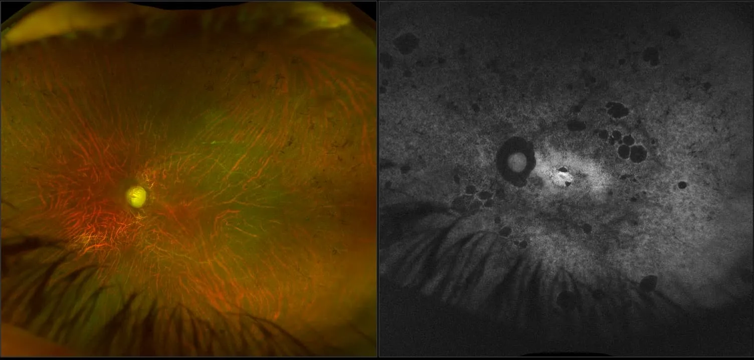
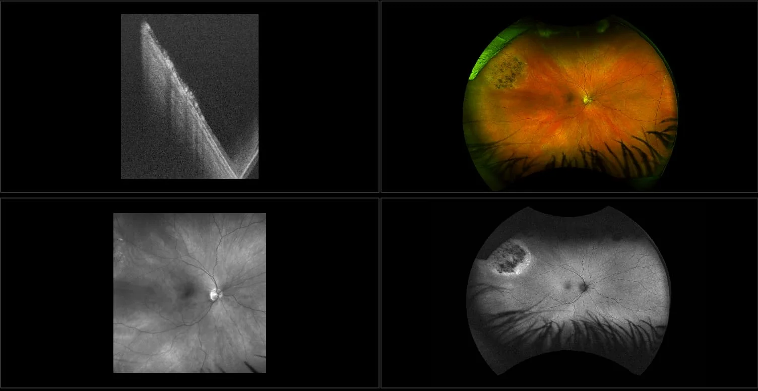
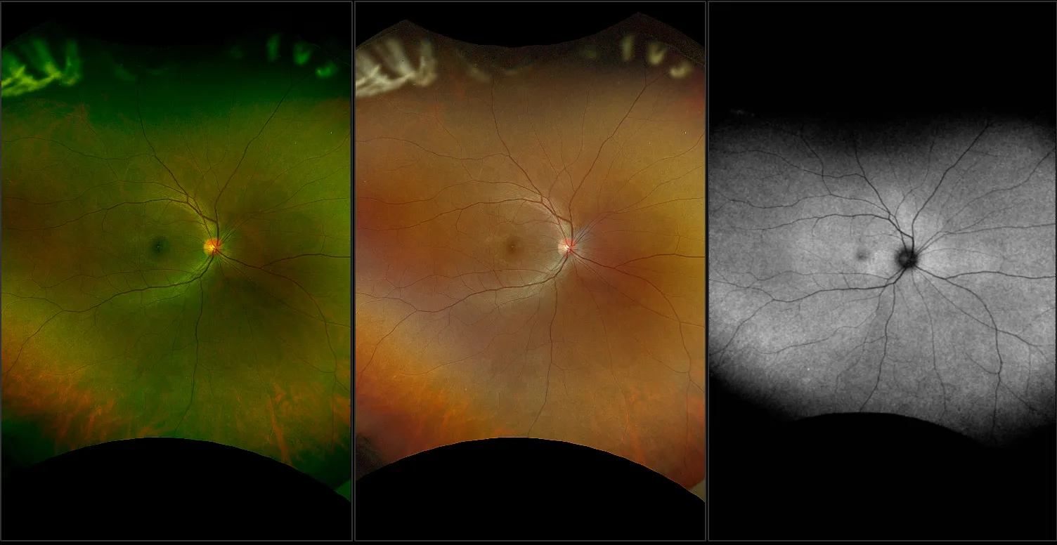
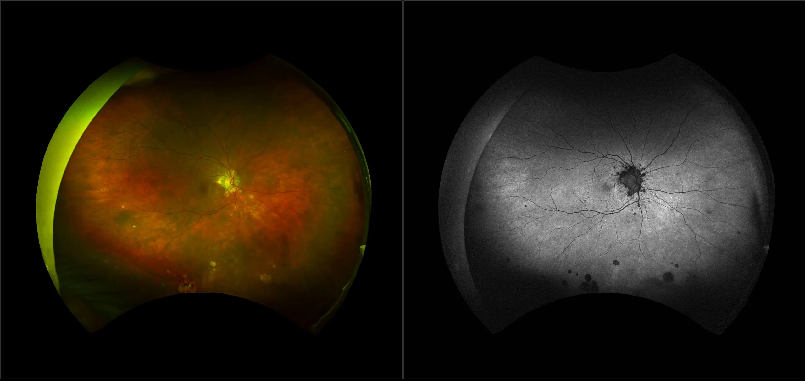
What: Uveitis is an inflammation of any of the structures of the uvea: iris, ciliary body, or choroid. Uveitis is often captured using multimodal imaging. This case demonstrates how those imaging modes work together to help confirm a patient’s disease.
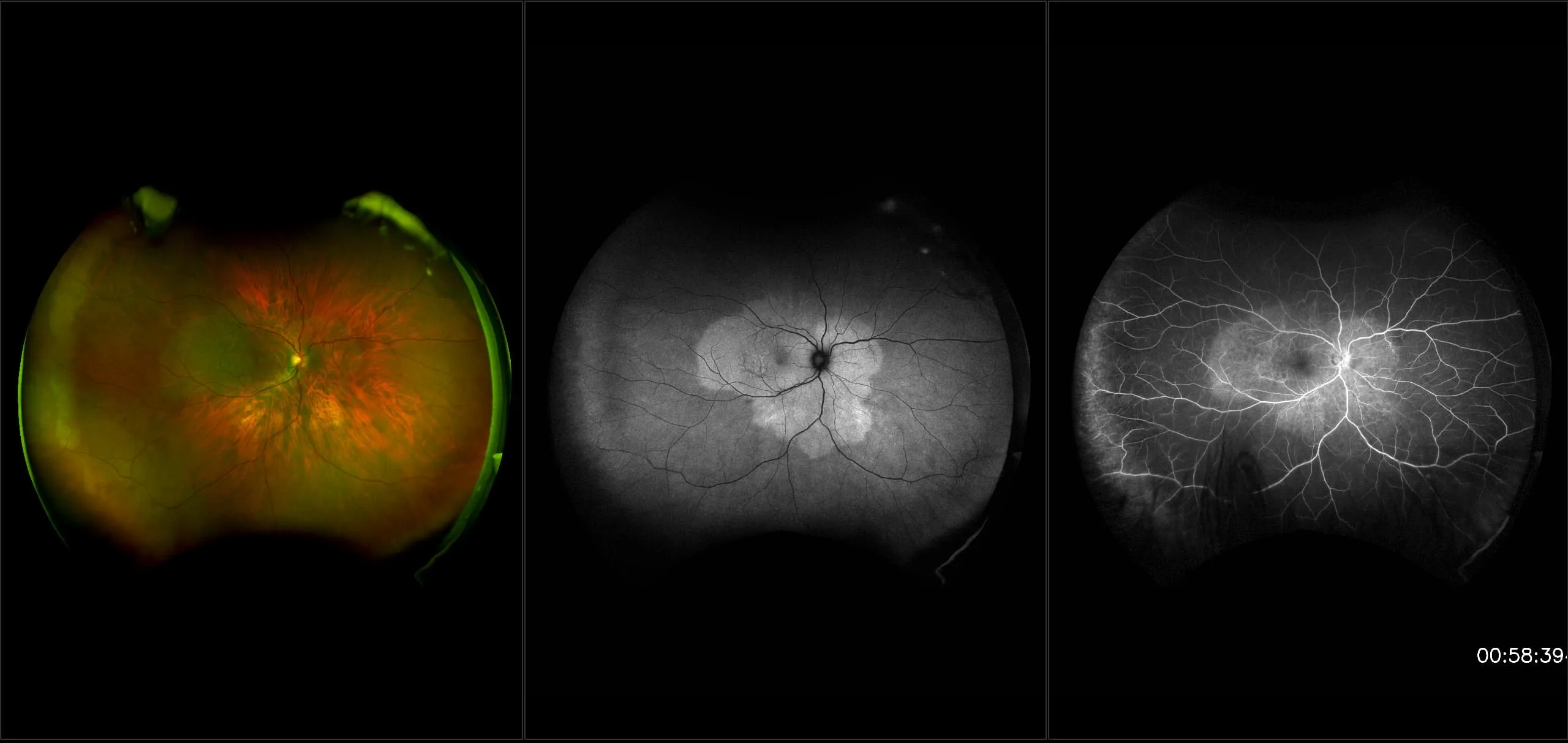
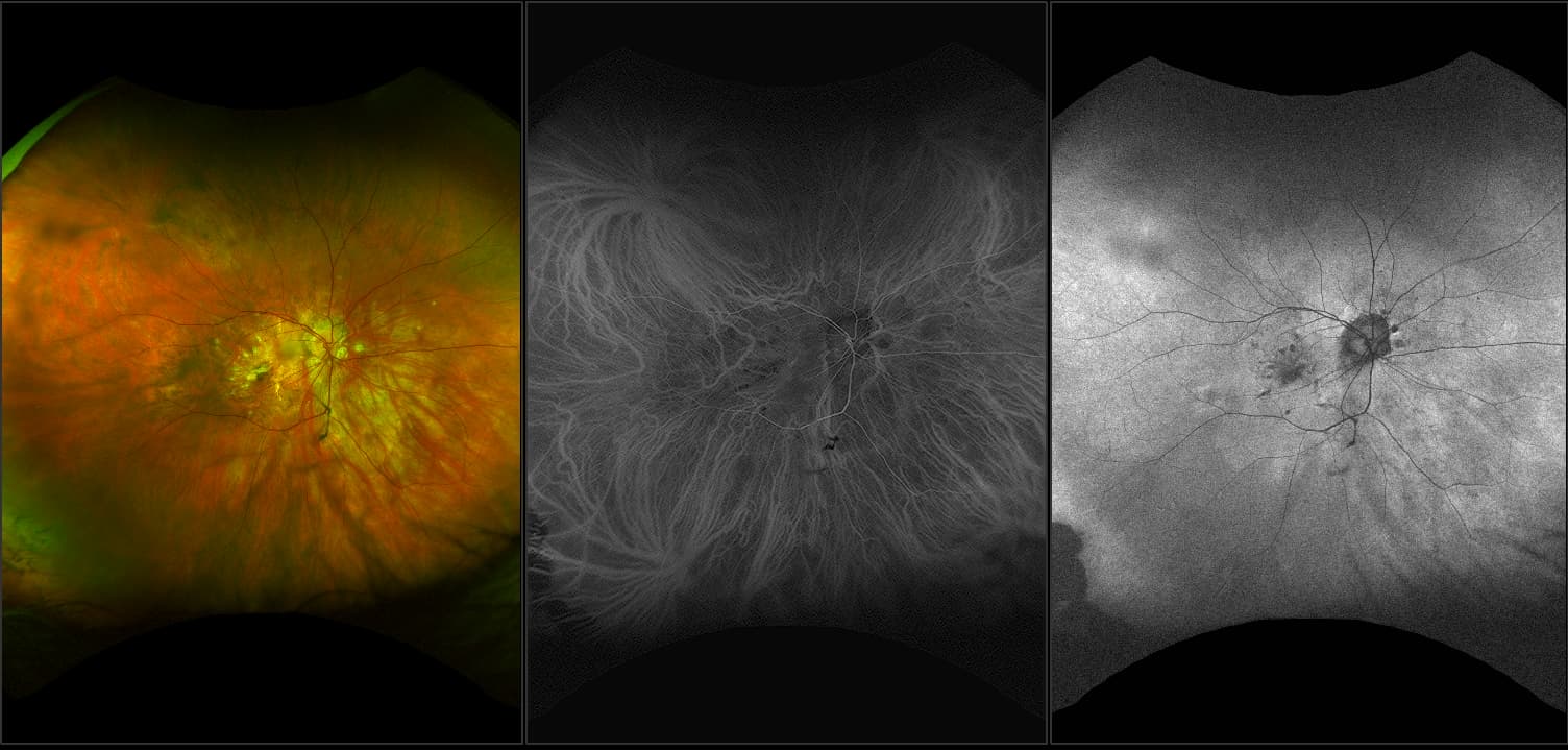
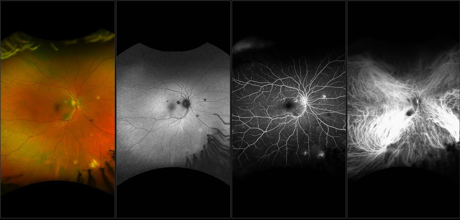
Why: A patient with uveitis was captured using optomap multimodal imaging. Images show inflammatory foci typical of uveitis in the mid to far periphery. These lesions correspond to each other on multimodal imaging. Interweave FA/ICG is often used when capturing uveitis. FA imaging may show pinpoint lesions and the ICG may help to confirm disease if lesions are seen in the ICG images.
In the picture: One study found that 59% of uveitis cases had peripheral findings on optomap icg. Another study found that adding UWF imaging changed treatment in uveitis patients up to 48% of the time.
References:
- Klufas. Feasibility and Clinical Utility of Ultra-widefield Indocyanine Green Angiography. Retina. 2014.
- Wide-field Retinal Imaging in the Management of Noninfectious Posterior Uveitis. American Journal of Ophthalmology. 2012
- Campbell et al. Wide-field retinal imaging in the management of non-infectious posterior uveitis. American Journal of Ophthalmology, 2012.