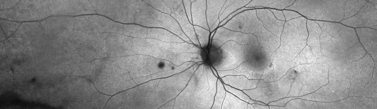
optomap af for Hydroxychloroquine Screening
Up to 70% of eyes with hydroxychloroquine (HCQ) retinopathy have findings captured on optomap af outside the central pole.1
American Academy of Ophthalmology Recommendations on Screening for Hydroxychloroquine Retinopathy
American Academy of Ophthalmology has updated their screening guidelines to encourage the use of wide-pattern AF as optomap af supports recognition of atypical toxicity patterns, which can be challenging to recognize with conventional FAF and horizontal OCT scans.2 Hydroxychloroquine (HCQ) is a frequently prescribed medication used for the management of rheumatoid arthritis, systemic lupus erythematosus (SLE), sarcoidosis, related autoinflammatory and dermatologic conditions.3 Irreversible retinal toxicity secondary to HCQ therapy has a well documented risk profile, which increases with cumulative dose and duration of therapy.4,5 The retina may remain normal for many years, but there is an unpredictable point at which evidence of toxicity can appear and progress relatively quickly to retinopathy.3 With 5 years of HCQ use, the risk of retinal toxicity is 7.5%, rising to almost 20% after 20 years of therapy, underlining the importance of careful, effective screening.6
After nearly a decade, the American Academy of Ophthalmology (AAO) published updated screening guidelines for HCQ retinopathy recommending fundus examination, OCT and ultra-widefield (UWF) AF at the start of HCQ therapy.3
Patterns of disease have been shown to differ based on heritage. Patients of European descent as well as those from India, Pakistan and the Middle East often display damage in a parafoveal distribution. Whereas patients of East Asian descent tend to show initial damage in a more peripheral extramacular distribution. However, these patterns are not exclusive and screening is recommended to observe for both patterns.3
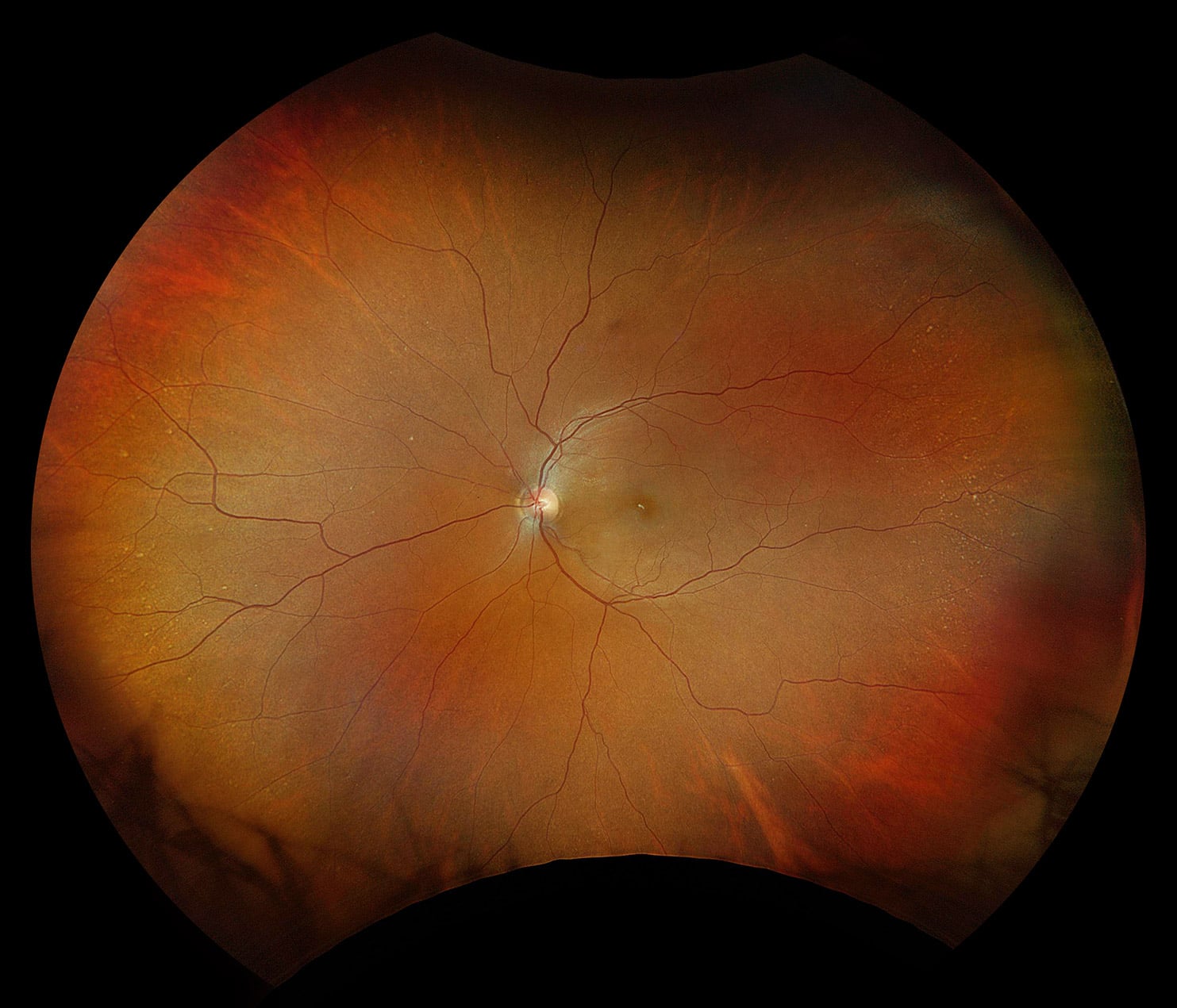
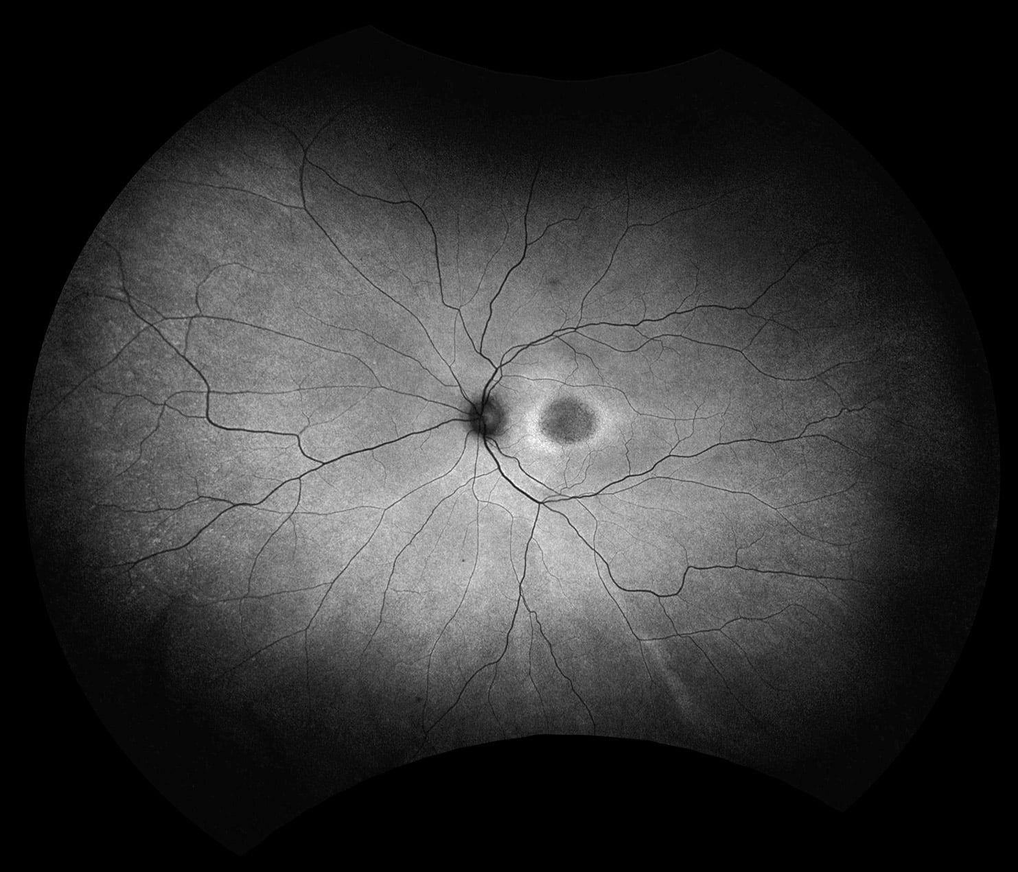
optomap color rgb and optomap green af
Why Optos?
Optos offers the only single capture, 200° UWF AF image meeting AAO’s updated screening recommendations. 70% of eyes with HCQ retinopathy have abnormal AF findings captured on optomap af outside the field of view of conventional AF.1 Additionally, 15% of patients exhibit atypical presentations of HCQ retinal toxicity including far peripheral dominant changes,2 and in 9% of eyes differences are revealed in detecting retinopathy between optomap green af and conventional AF.1
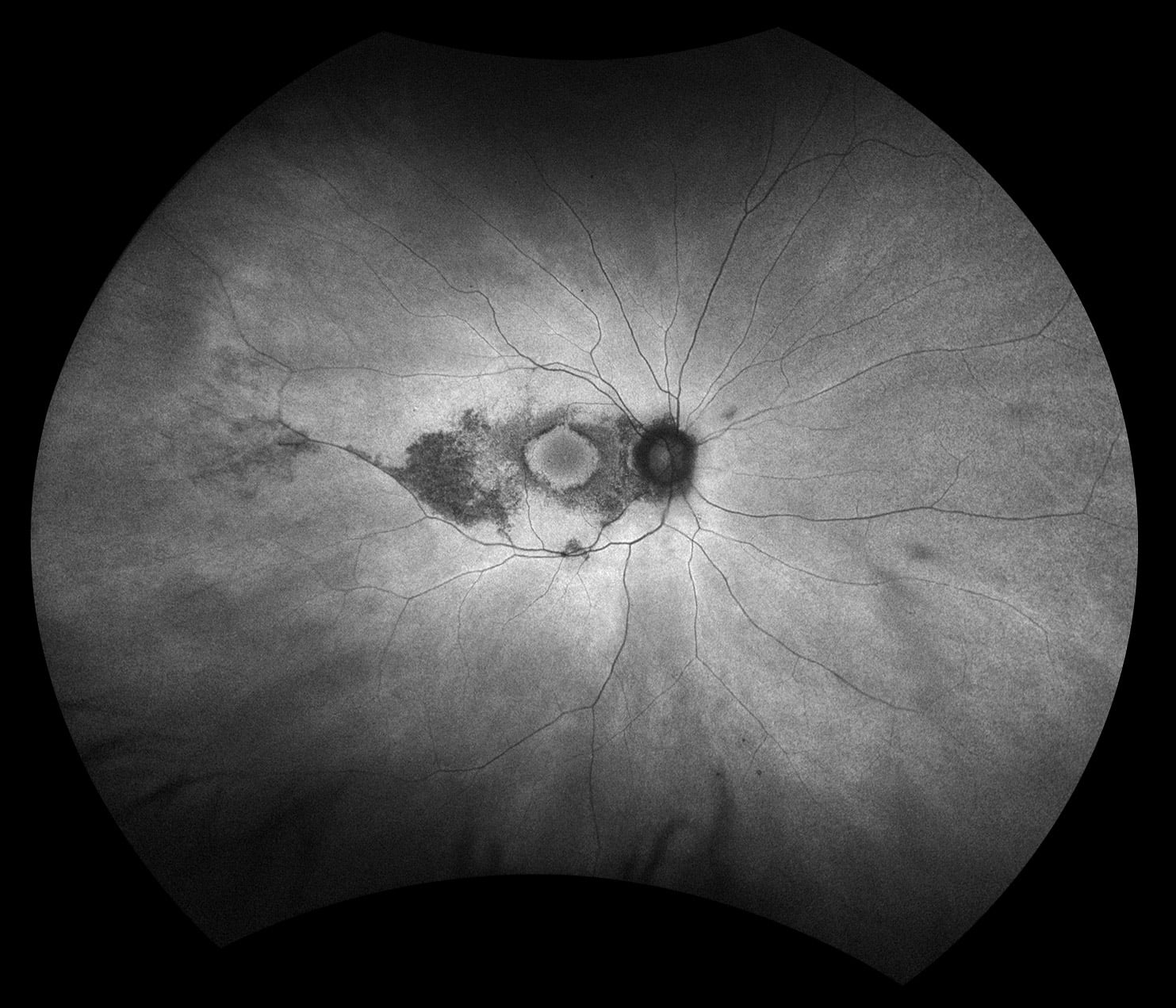
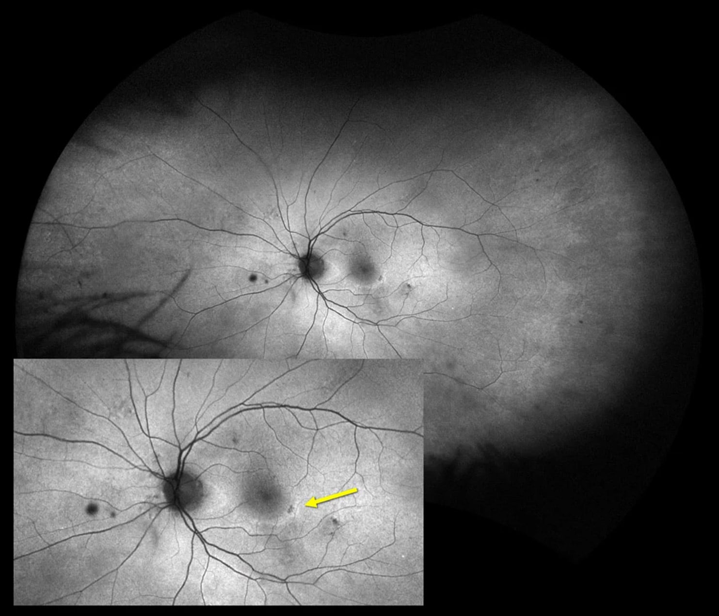
optomap green af and optomap green af
The earliest signs of HCQ toxicity for either pattern of involvement are areas of increased autofluorescence, most often in the inferotemporal quadrant.1
OptosAdvanceTM allows for efficient image review7 and facilitates monitoring for progression over time, which may be suitable for comparing baseline HCQ screening examination to future follow-ups. Additionally, OptosAdvance is a PACS system and can be leveraged to store and review non-Optos examination methods, such as visual field reports. OptosAdvance may also be beneficial for education, using tools such as 3D wrap to counsel patients, caregivers and referring physicians on related findings.
Unfortunately, compliance with HCQ screening recommendations is fair at best.3 Poor adherence to these guidelines, in particular delayed initial monitoring, may be associated with late diagnosis of HCQ retinopathy.8 optomap has been shown to increase compliance with annual screening guidelines and may therefore support timely diagnosis.9
Reference Materials
Recent Papers Utilizing Optos UWF for Hydroxychloroquine
References
1. Ahn, S. J., Joung, J., & Lee, B. R. (2020). Evaluation of Hydroxychloroquine Retinopathy Using Ultra-Widefield Fundus Autofluorescence: Peripheral Findings in the Retinopathy. American journal of ophthalmology, 209, 35–44. https://doi.org/10.1016/j.ajo.2019.09.008
2. Lee, J. M., Kwon, H. Y., & Ahn, S. J. (2024). Atypical Presentations of Hydroxychloroquine Retinopathy: A Case Series Study. Journal of clinical medicine, 13(12), 3411. https://doi.org/10.3390/jcm13123411
3. Marmor MF, Ahn SJ, Ehlers JP, Melles RB, Mieler WF, Sarraf D, Yusuf IH, for the American Academy of Ophthalmology, Special Report: Recommendations on Screening for Hydroxychloroquine Retinopathy (2026 Revision), Ophthalmology (2025), doi: https://doi.org/10.1016/j.ophtha.2025.11.001.
4. Yusuf, I. H., Charbel Issa, P., & Ahn, S. J. (2023). Hydroxychloroquine-induced Retinal Toxicity. Frontiers in pharmacology, 14, 1196783. https://doi.org/10.3389/fphar.2023.1196783
5. Melles, R. B., Jorge, A. M., Marmor, M. F., Zhou, B., Conell, C., Niu, J., McCormick, N., Zhang, Y., & Choi, H. K. (2023). Hydroxychloroquine Dose and Risk for Incident Retinopathy : A Cohort Study. Annals of internal medicine, 176(2), 166–173. https://doi.org/10.7326/M22-2453
6. Melles, R. B., & Marmor, M. F. (2014). The risk of toxic retinopathy in patients on long-term hydroxychloroquine therapy. JAMA ophthalmology, 132(12), 1453–1460. https://doi.org/10.1001/jamaophthalmol.2014.3459
7. Silva, P. S. et al. (2016). Identification of Diabetic Retinopathy and Ungradable Image Rate with Ultrawide Field Imaging in a National Teleophthalmology Program. Ophthalmology, 123(6), 1360–1367. https://doi.org/10.1016/j.ophtha.2016.01.043
8. Ahn, S. J., & Kim, J. H. (2024). SCREENING PRACTICES AND LATE DIAGNOSIS OF HYDROXYCHLOROQUINE RETINOPATHY IN ASIAN PATIENTS. Retina, 44(7), 1251–1259. https://doi.org/10.1097/IAE.0000000000004086
9. Lin, C. C., Li, A. S., Ma, H., Lin, X. M., Olivares, M. Z., Haubrich, A., Sanislo, S., & Do, D. V. (2021). SUCCESSFUL INTERVENTIONS TO IMPROVE EFFICIENCY AND REDUCE PATIENT VISIT DURATION IN A RETINA PRACTICE. Retina, 41(10), 2157–2162. https://doi.org/10.1097/IAE.0000000000003169