You are now leaving the Global site.

Ok Cancel
optos
  • Products
  • Patients
  • Providers
  • About Us
  • Contact
  • Customers
  • Language Menu
    English Deutsch Français

optomap® Recognizing Pathology

This material is designed as a searchable reference resource to support clinical decision-making. The information contained here should be used as general guidance when viewing optomap and OCT images from Optos devices. The differential diagnosis should be made under the direction of the responsible physician. These images were taken on the latest ultra-widefield optomap devices.
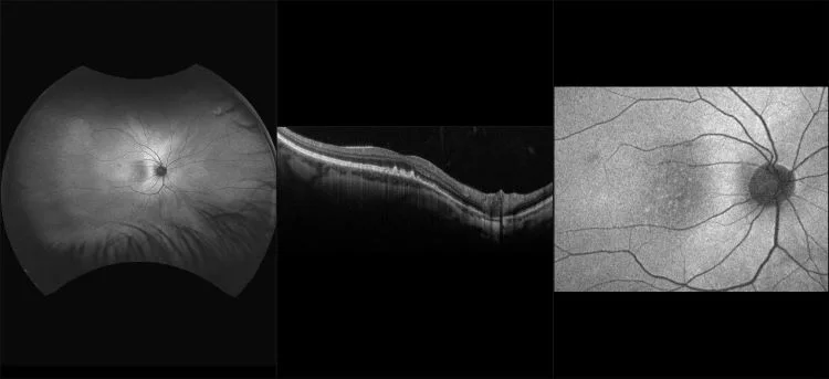
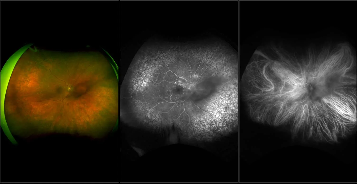
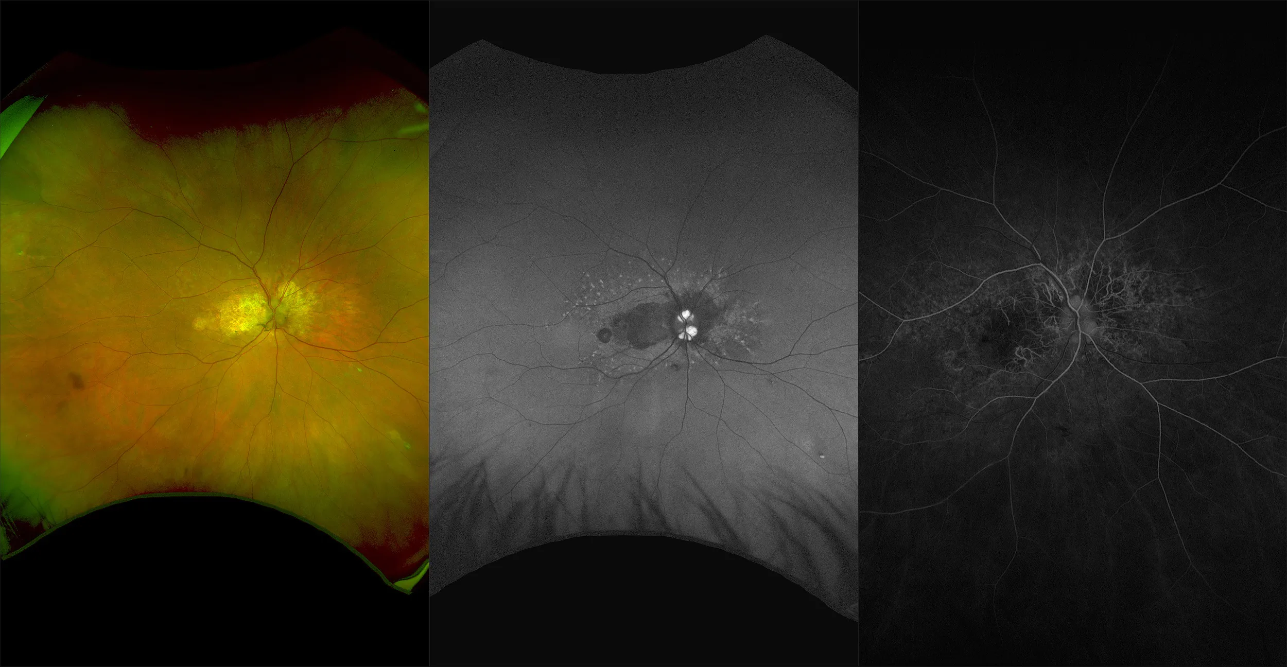
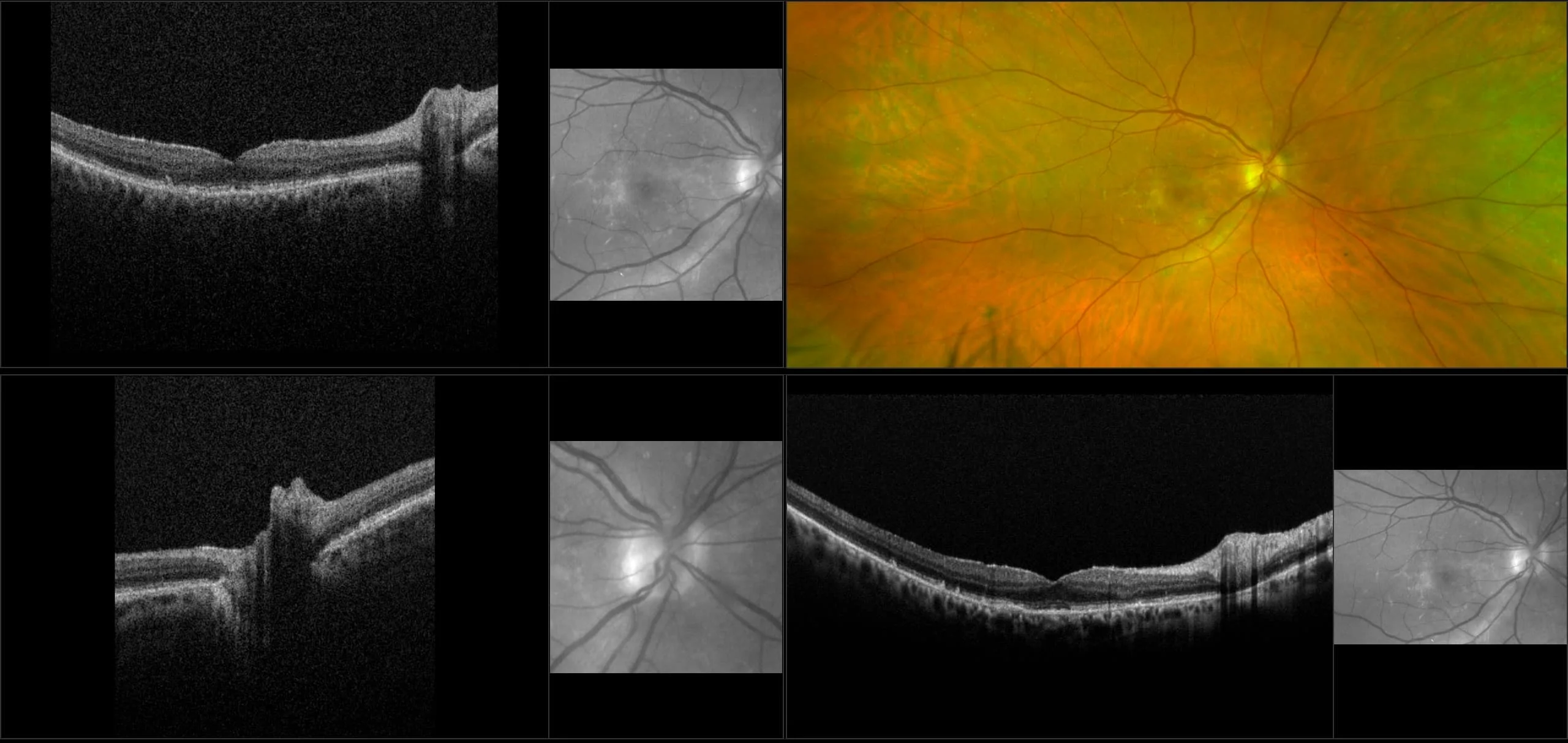
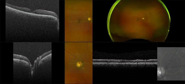
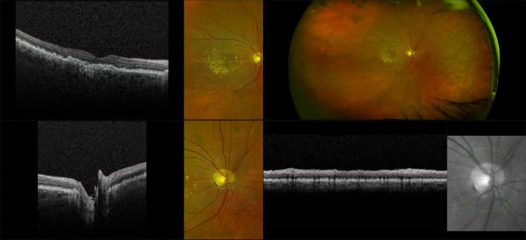
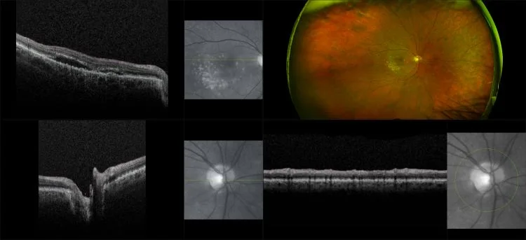
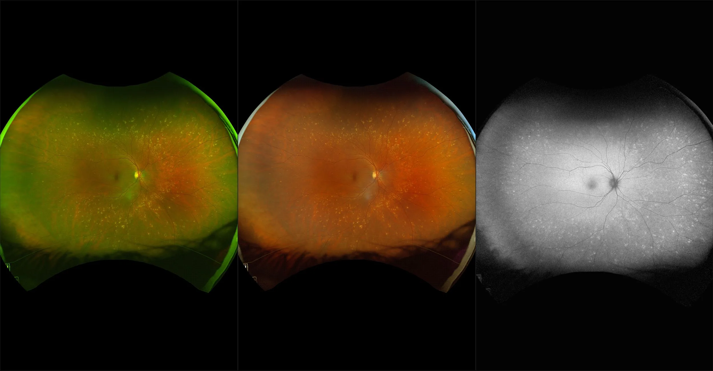
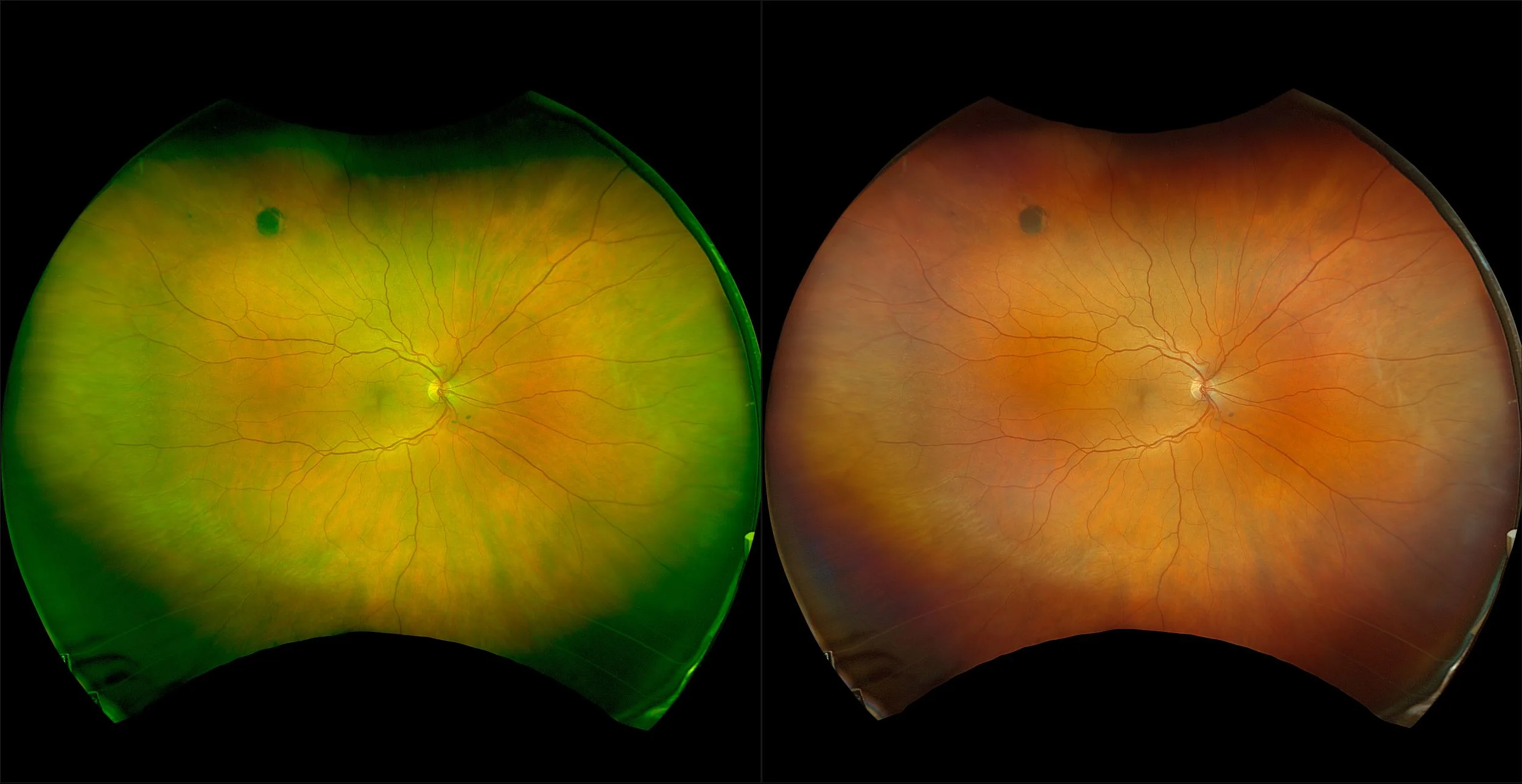
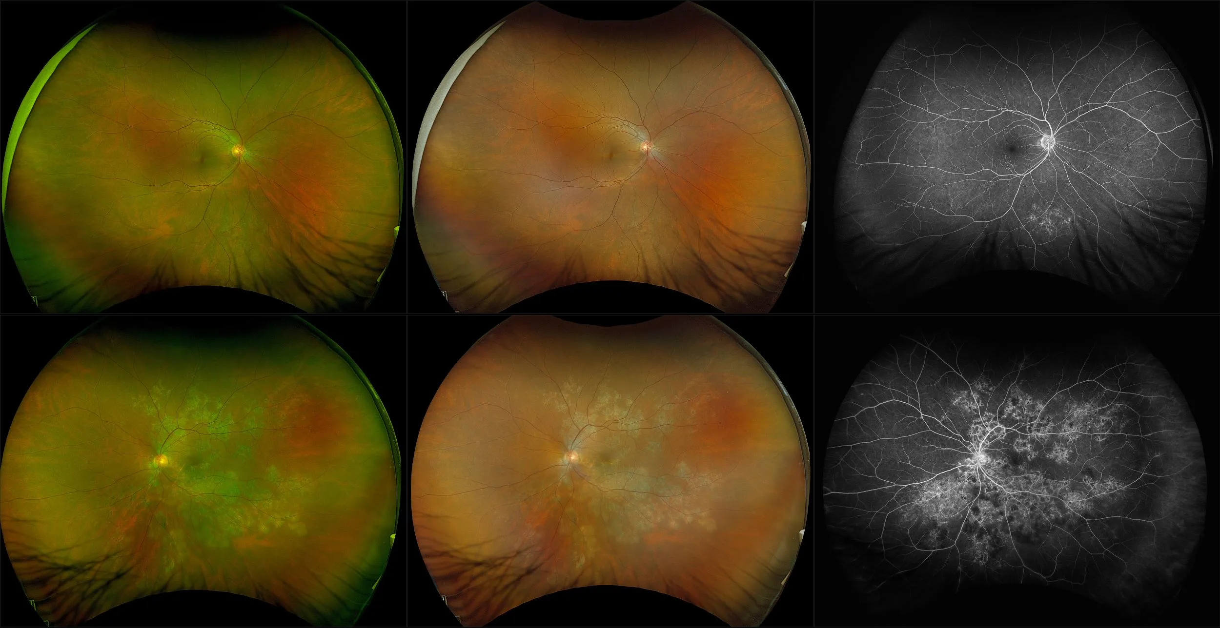
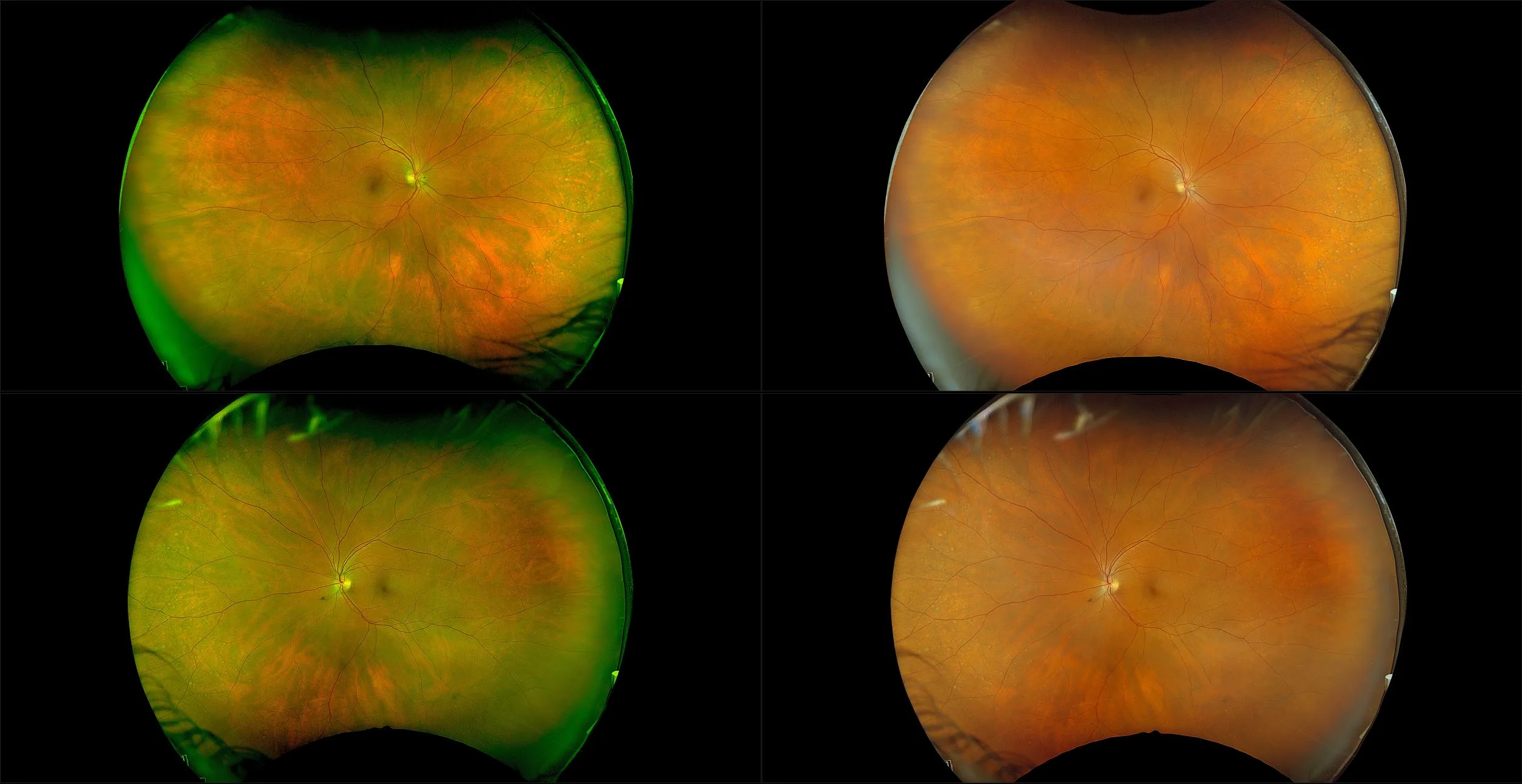
Pathology Definitions
Help and Support

Drusen

Lipid and protein-rich deposits at the level of Bruch’s membrane below the RPE. Small, hard drusen are common with age (present in the majority of individuals over the age of 60), but larger drusen are a hallmark feature of age-related macular degeneration.

How can we help?

If you have questions regarding the cases, images, or pathologies, or if you would like further information about our ultra-widefield retinal imaging technology, please complete the form below.
I want to receive email updates about Optos products and services
<p>You are now leaving the Global site.</p> Ok Cancel
  • Blog
  • Cookie Policy
  • Careers
  • Distributor Portal
  • Manage Preferences
  • Media
  • News
  • Privacy
  • Registered in Scotland
  • UK Modern Slavery Act

© 2023 Optos. All rights reserved. Company Registration Number SC139953