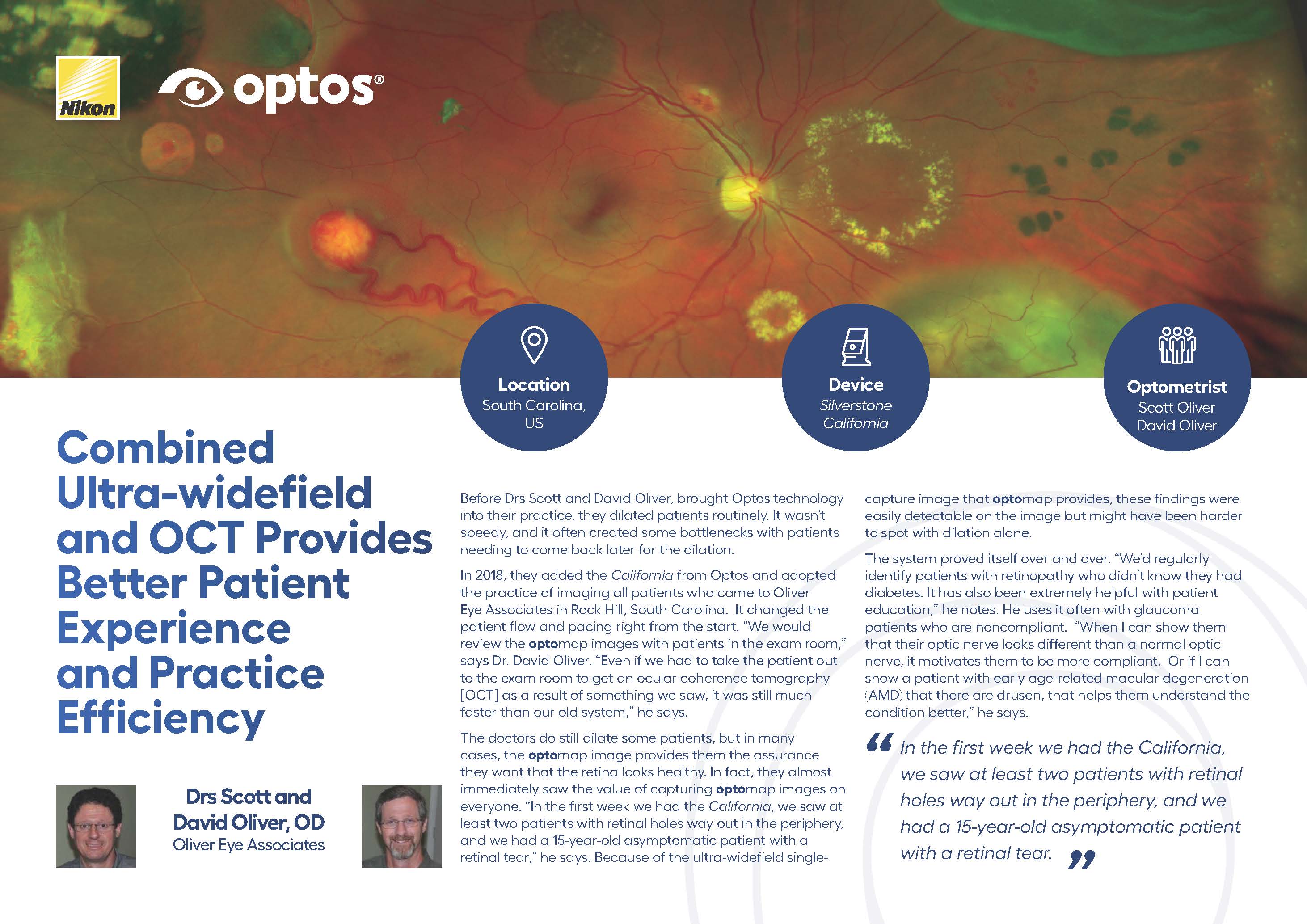
Optometrists Scott & David Oliver brought Optos technology into their practice in 2018, with the purchase of California. They immediately implemented imaging all patients who came to their practice. From the start, it changed the patient flow and pacing. “We would review the optomap images with patients in the exam room,” says Dr. David Oliver. “Even if we had to take the patient out to the exam room to get an OCT as a result of something we saw, it was still much faster than our old system,” he says.
The optomap image provides them the assurance they want that the retina looks healthy. In fact, they almost immediately saw the value of performing optomap images on everyone. “In the first week we had California, we saw at least two patients with retinal holes way out in the periphery, and we had a 15-year-old asymptomatic patient with a retinal tear,” he says. Because of the ultra-widefield single-capture image that optomap provides, these findings were easily detectable on the image but might have been harder to spot with dilation alone.
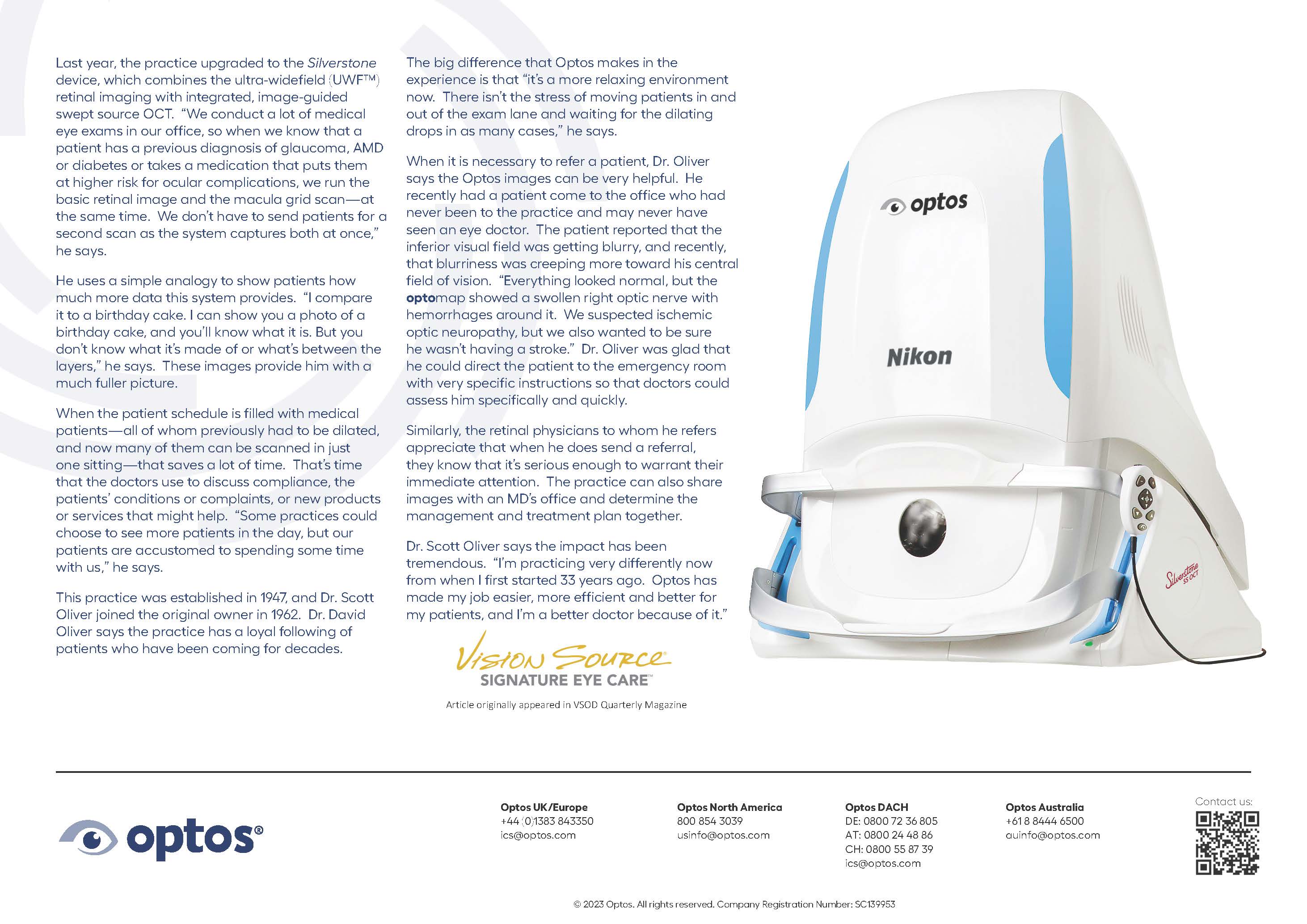
Last year, the practice upgraded to the Silverstone device, which combines ultra-widefield retinal imaging with integrated, image-guided swept-source OCT.
He uses a simple analogy to show patients how much more data this system provides. “I compare it to a birthday cake. I can show you a photo of a birthday cake, and you’ll know what it is. But you don’t know what it’s made of or what’s between the layers,” he says. These images provide him with a much fuller picture.
When it is necessary to refer a patient, Dr. Oliver says the optomap images can be very helpful. He recently had a patient come to the office who had never been to the practice and may never have seen an eye doctor. The patient reported that the inferior visual field was getting blurry, and recently, that blurriness was creeping more toward his central field of vision. “Everything looked normal, but the optomap showed a swollen right optic nerve with hemorrhages around it. We suspected ischemic optic neuropathy, but we also wanted to be sure he wasn’t having a stroke.” Dr. Oliver was glad that he could direct the patient to the emergency room with very specific instructions so that doctors could assess him specifically and quickly.
Dr. Scott Oliver says the impact has been tremendous. “I’m practicing very differently now from when I first started 33 years ago. Optos has made my job easier, more efficient, and better for my patients, and I’m a better doctor because of it.”
Read Drs.. Oliver full testimonial for more firsthand knowledge of the benefits of optomap imaging with integrated OCT, in optometric practice.