Diabetic retinopathy is one of the leading causes of vision loss and blindness around the globe, and affects approximately one-third of people with diabetes. Because the number of people with diabetes is expected to rise to more than 438 million worldwide by 2030, it can be assumed that the prevalence of diabetic retinopathy can be expected to rise, as well.
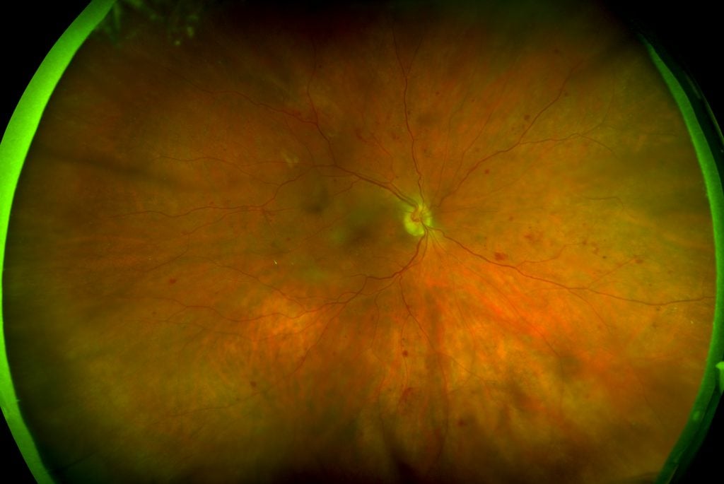
In this optomap image, you can see that the leaky vessels denoting the presence of diabetic retinopathy, often seen far out on the edges, where they may go undetected using traditional retinal imaging methods.
For individuals with diabetes, certain risk factors can be reduced by focusing on key elements such as education, early detection, and early treatment.
What Is Diabetic Retinopathy?
Diabetes impairs the body’s ability to control blood sugar. Because sugar can promote inflammation, a person with diabetes may experience inflammatory damage to various tissues within their body, including the retina, which is located on the inside back of your eye. High blood sugar causes damage to the blood vessels in the retina, where the small blood vessels that supply blood to their retinal tissue become leaky and irritated. Over time, this can lead to complete vision loss if not treated properly.
Why Is Early Diagnosis of Diabetic Retinopathy So Important?
The critical challenge with diabetic retinopathy is that retinal damage often begins long before symptoms ever develop. As such, individuals with diabetes could be heading toward vision loss without even knowing.
In addition, it’s not always possible to reverse tissue damage and/or vision loss that occurs due to diabetic retinopathy. However, with appropriate diagnosis and treatment, eyecare professionals may be able to help prevent further deterioration. For this reason, early detection of diabetic retinopathy is critical.
The Role of optomap® in the Diagnosis of Diabetic Retinopathy
Eyecare professionals can greatly enhance their ability to provide early detection of diabetic retinopathy and other eye conditions with the use of advanced ultra-widefield (UWF™) diagnostic technology like optomap®.
optomap is specifically designed to provide an ultra-widefield image of the retina, and it is the only technology that captures a 200 degree image of the retina in a single scan and in less than ½-second. Because optomap can image so far out in the periphery, where the damage from diabetic retinopathy often begins, it allows eyecare professionals to get a clear look at the health of the retina and determine if there are any early warning signs of diabetic retinopathy. optomap can also be used to confirm a diagnosis, allowing eyecare professionals to initiate a plan of care as soon as possible.
What Is the Prognosis for People with Diabetic Retinopathy?
Factors that influence an eyecare professionals clinical decision-making about the best treatment options will include considerations such as overall health status; how well the underlying diabetes is controlled; the severity, type, and duration of any symptoms; and age.
Prognostic research suggests great variability in outcomes for people with diabetic retinopathy. However, the majority of patients who receive the gold standard of care for diabetic retinopathy can generally expect to see some improvement in their eyesight following treatment, if not a slowing of the disease process altogether.
The bottom line is that people with diabetes are best served by eyecare professionals who use optomap in order to improve the chances of early detection and early treatment of any diabetes-related eye diseases. As always, it is critical to work closely your eyecare provider and primary care doctor to ensure everything is being done to manage the ancillary impacts that diabetes can have on your health.
To find an eyecare professional near you who uses optomap technology, visit: www.optomap.com.
Source: