optomap® Recognizing Pathology
This material is designed as a searchable reference resource to support clinical decision-making. The information contained here should be used as general guidance when viewing optomap and OCT images from Optos devices. The differential diagnosis should be made under the direction of the responsible physician. These images were taken on the latest ultra-widefield optomap devices.
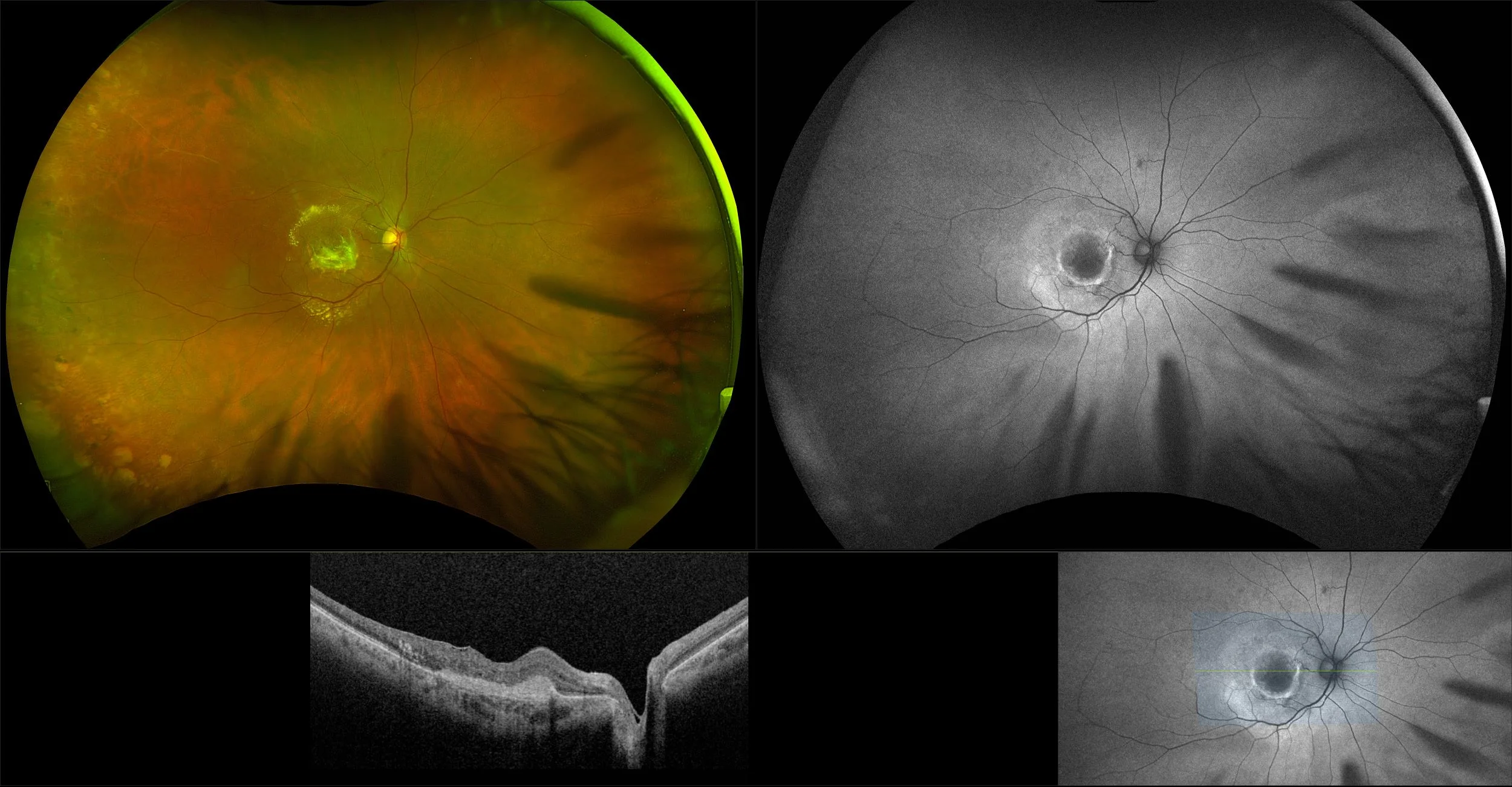
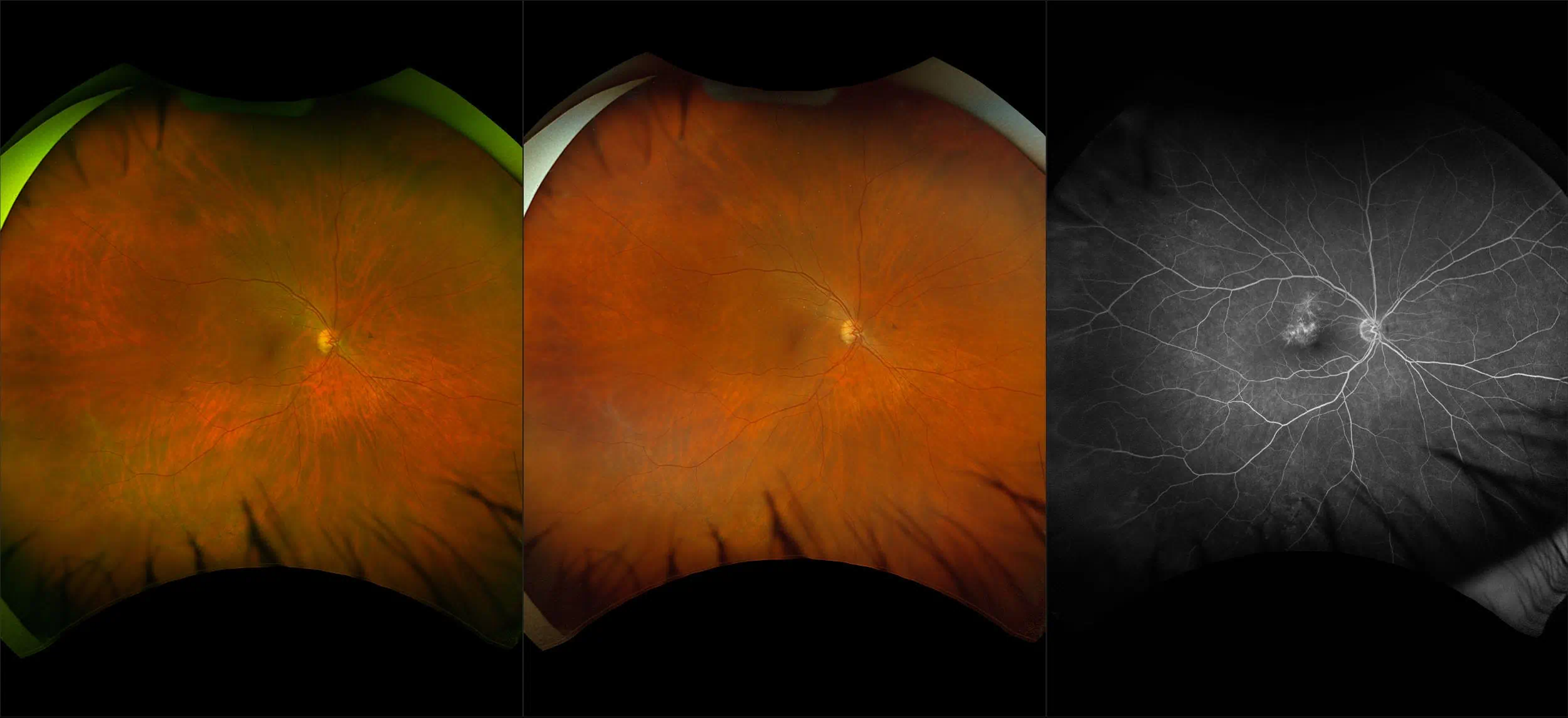
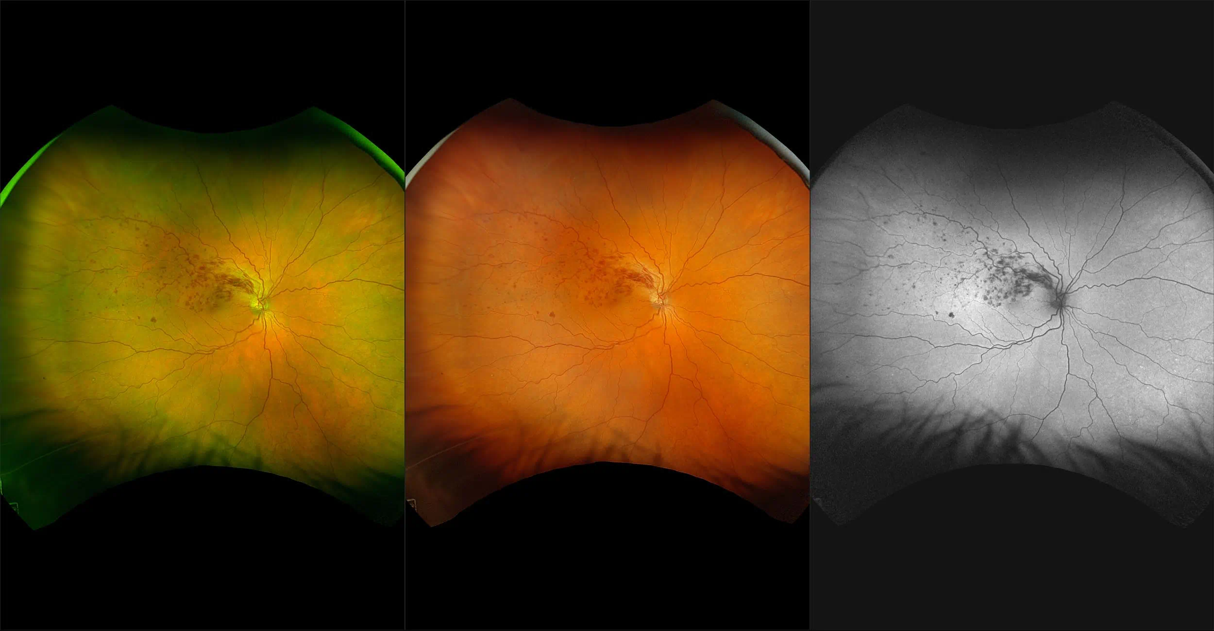
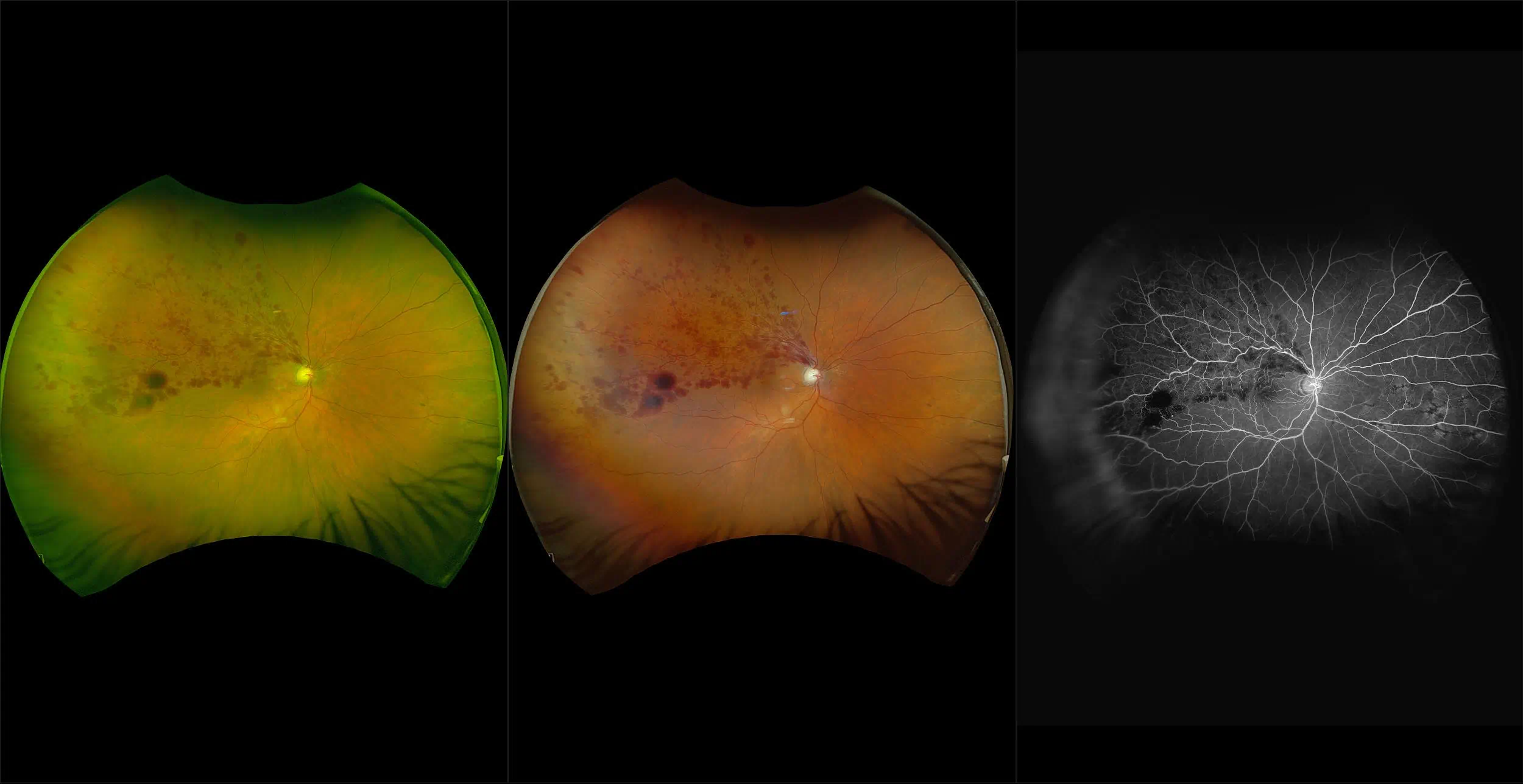
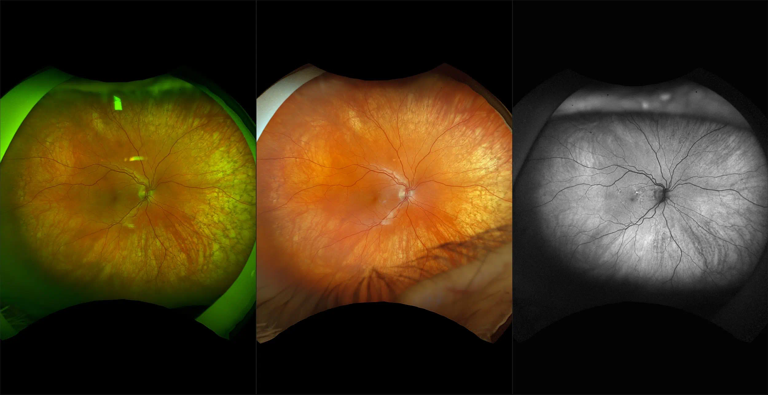
Branch Retinal Vein Occlusion (BRVO)
Branch retinal vein occlusion can be caused by thrombosis but is more likely to be produced by compression due to arteriolosclerosis. This occurs at arteriovenous crossing sites where there is a common adventitial sheath of each vessel. Marked A/V nicking usually precedes the occlusive process.