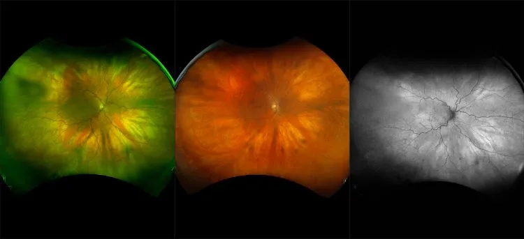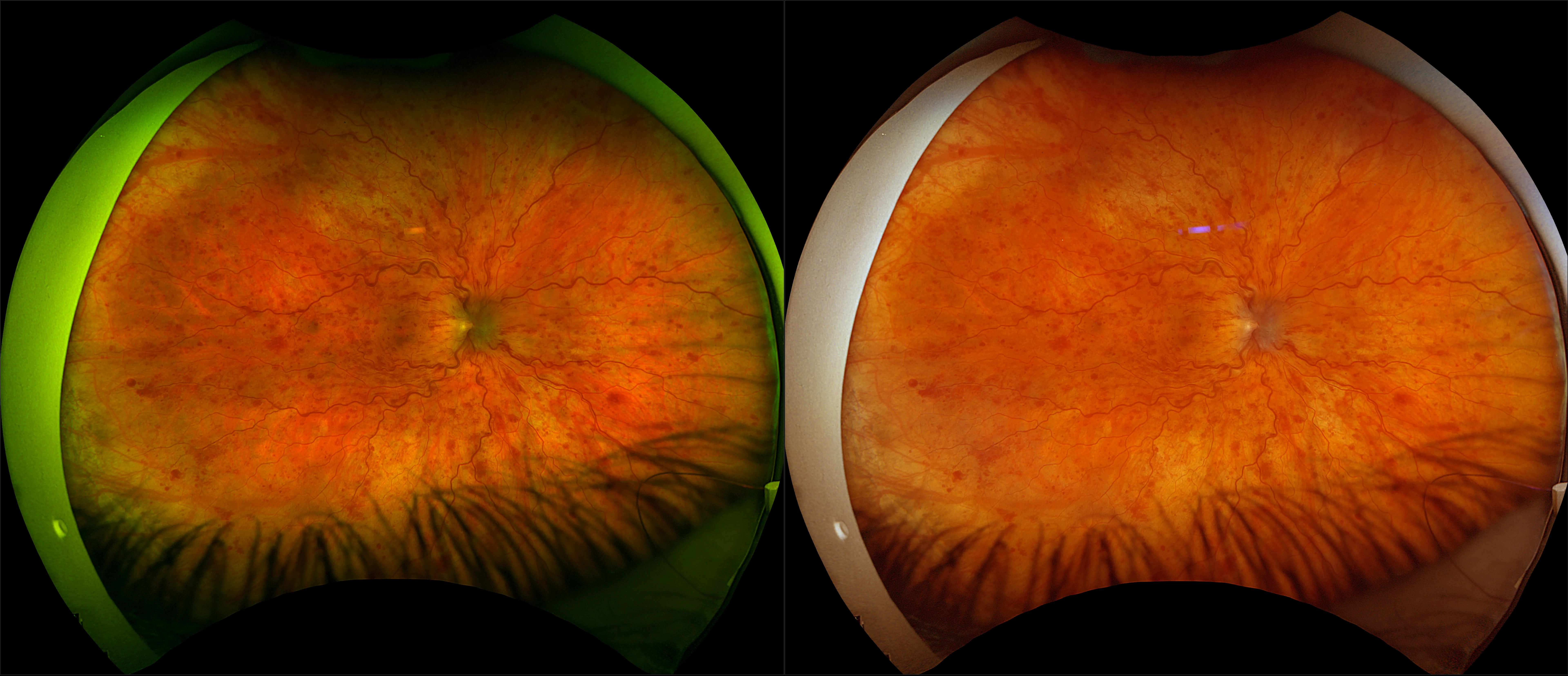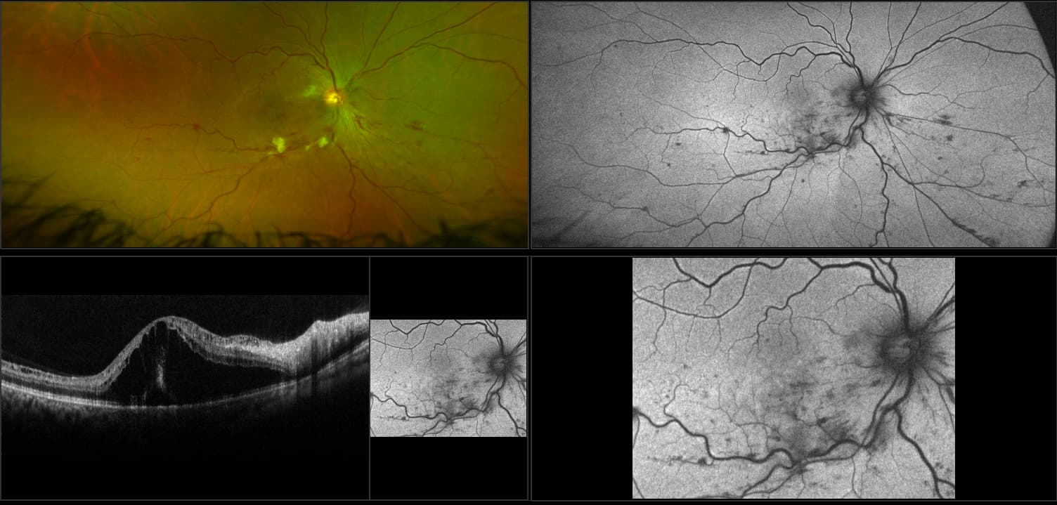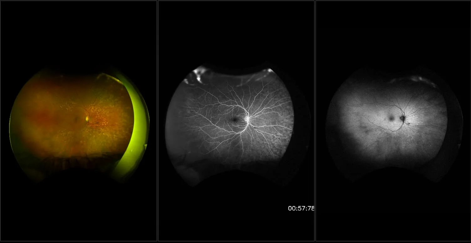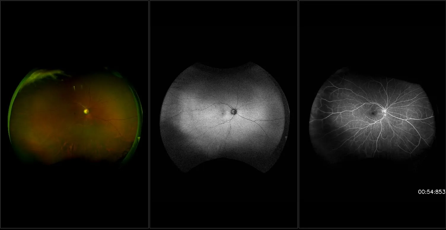
California - Hemi Retinal Vein Occlusion, RG, AF, FA
What: Retinal Vein Occlusion is a retinal vascular disorder that can involve the central retinal vein, a major branch of the central vein, or any part of a branch.
Why: On fluorescein angiography, retinal circulation is delayed showing areas of non-perfusion and the point of the occlusion can usually be visualized with angiography. Vessel leakage and staining can occur in the vessels extending to the region of occlusion. Cystoid macular edema (CME) can also be present and visual acuity may be greatly decreased.
In the picture: A patient of Dr. David Brown at Retina Consultants of Houston with a Hemi Retinal Vein Occlusion was imaged on the Optos. Images were superiorly steered to show the extent of the occlusion from the posterior pole to the far periphery. optomap color image shows the extent of the flamed shaped hemorrhages which is characteristic of RVO. Fluorescein angiography shows the vessel leakage and non-perfusion in the superior hemisphere of the retina. optomap fa allows for simultaneous capture of the central pole and periphery so that leakage can be seen across the retina at any given time. The leakage in the macula is indicative of macula edema.