Diagnostic & Educational Tools
Ultra-widefield retinal imaging technology lets you see more of the retina...easier. optomap images provide an unprecedented 200 degree view that enables practitioners to more effectively detect, monitor and promote patient health. The tools below will assist you in identifying pathology on optomap images.
Recognizing Pathology - The Complete optomap Clinical Image Library
Recognizing Pathology is designed as a searchable reference resource to support clinical decision making. The information contained here should be used as a general guideline only when viewing optomap images.
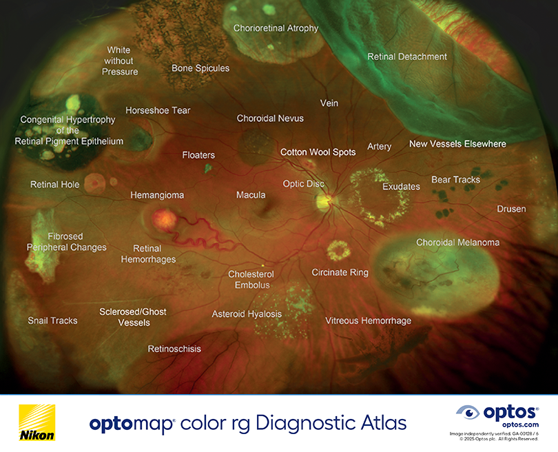
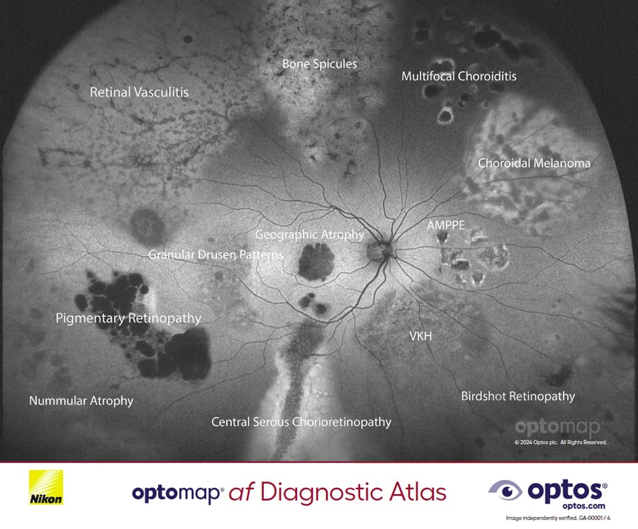
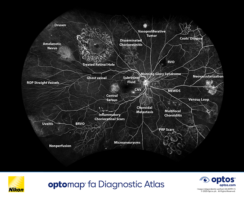
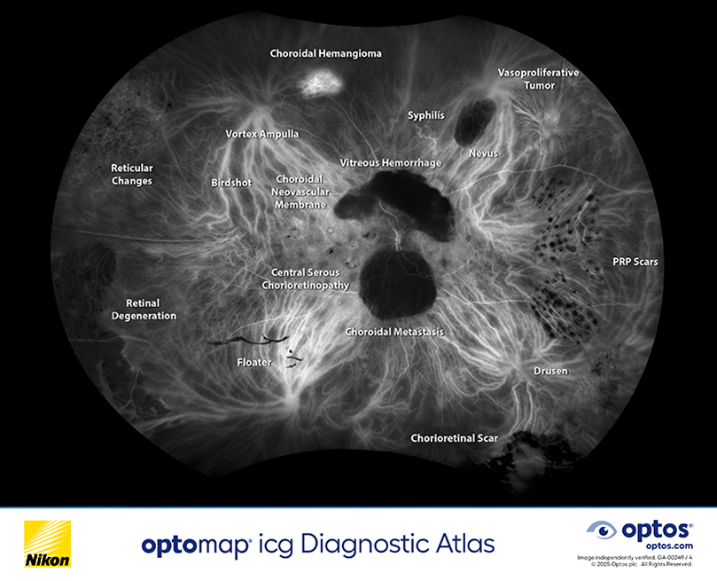
Downloadable Diagnostic Atlas PDFs
View, share, and download our popular diagnostic atlases. These tools are helpful for patient education regarding the types of disease and pathology that can occur on the retina.

Interactive Flipbooks (Color and OCT)
To further assist eyecare professionals in recognizing retinal pathology, we have expanded on our popular retinal atlas PDFs (above) and created a full handbook for all of our diagnostic atlases. Each pathology has expandable images and additional educational information.
View, share and download these tools.
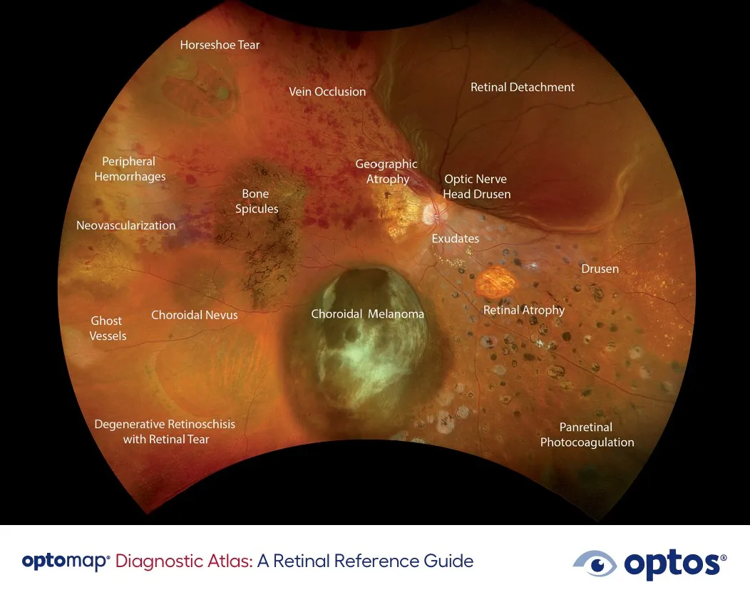
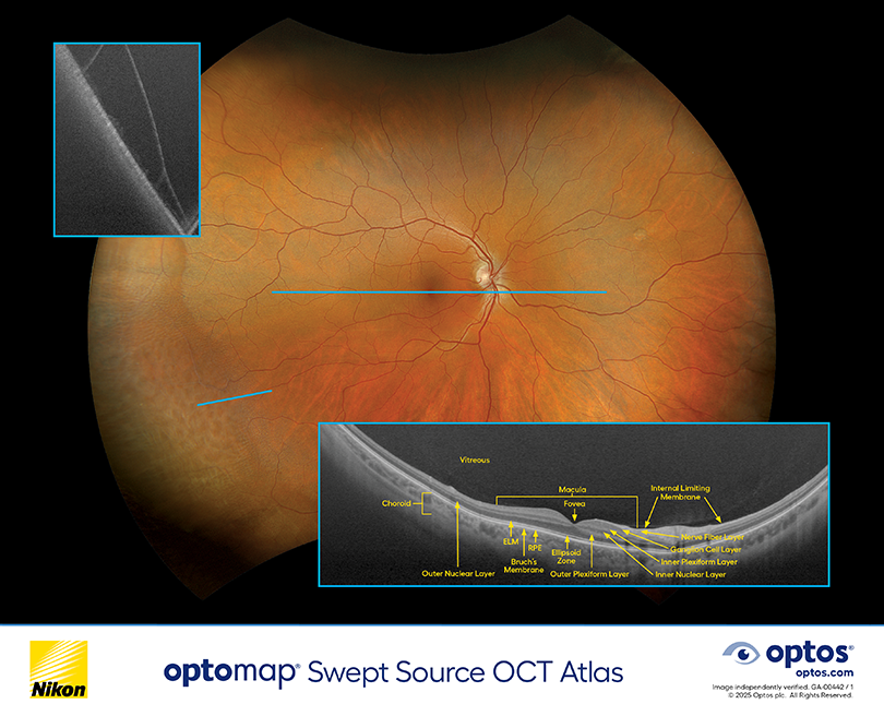
Diagnostic Atlas Booklet – optomap Core Modalities
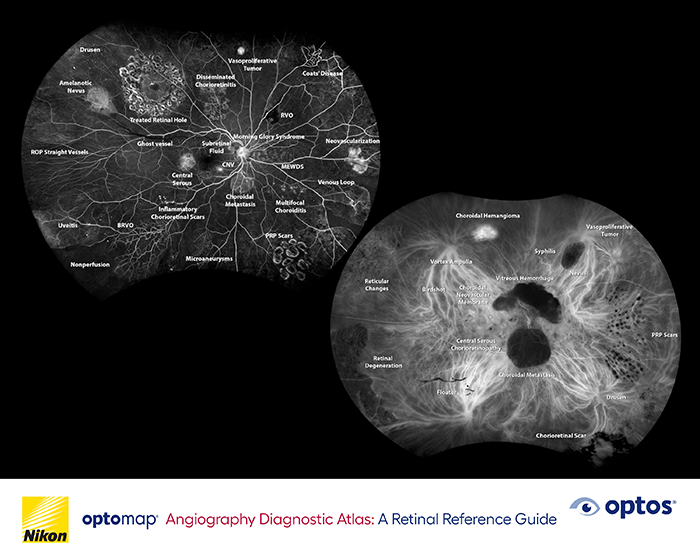
Diagnostic Atlas Booklet – optomap Angiography
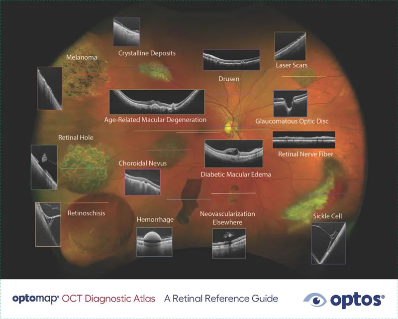
Diagnostic Atlas Booklet – optomap OCT
Videos
For a better understanding of the differences between optomap versus widefield or traditional retinal imaging devices, please watch the videos below.