Future proof your practice with Optos
In a climate of economic uncertainty, Optometrists with independent or small practices face several business challenges. Our research has revealed three key indicators impacting customer retention and financial growth.
3 key challenges optometrists face:
- Running costs & overheads
- Attracting new patients
- Retaining trained staff
An advancement in the standard of retinal eye care is being driven by the latest innovations in technology. Investment in your businesses today can result in improved clinical care for your patients into the future. The positive result of enhanced patient care with advanced retinal imaging can lead to an increase in customer loyalty and revenue growth. Optos technology is being embraced by optometrists who are using our range of devices to transform clinical eye care and grow customer and staff retention.
Invest in your practice with Optos technology
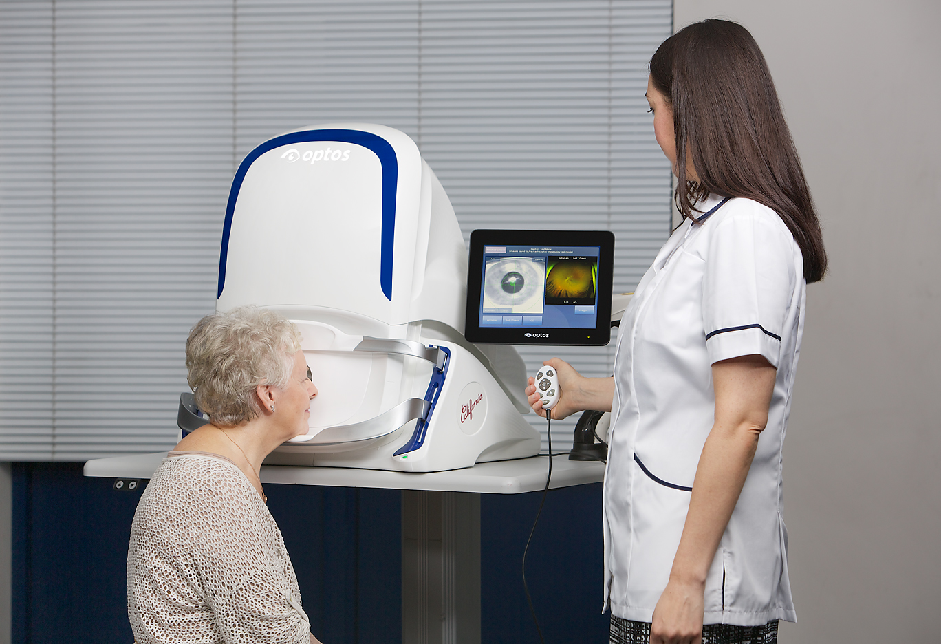
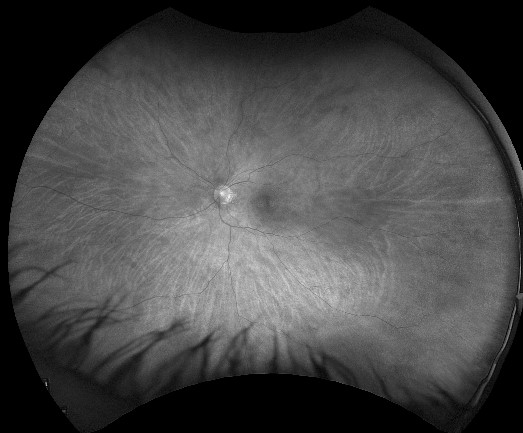
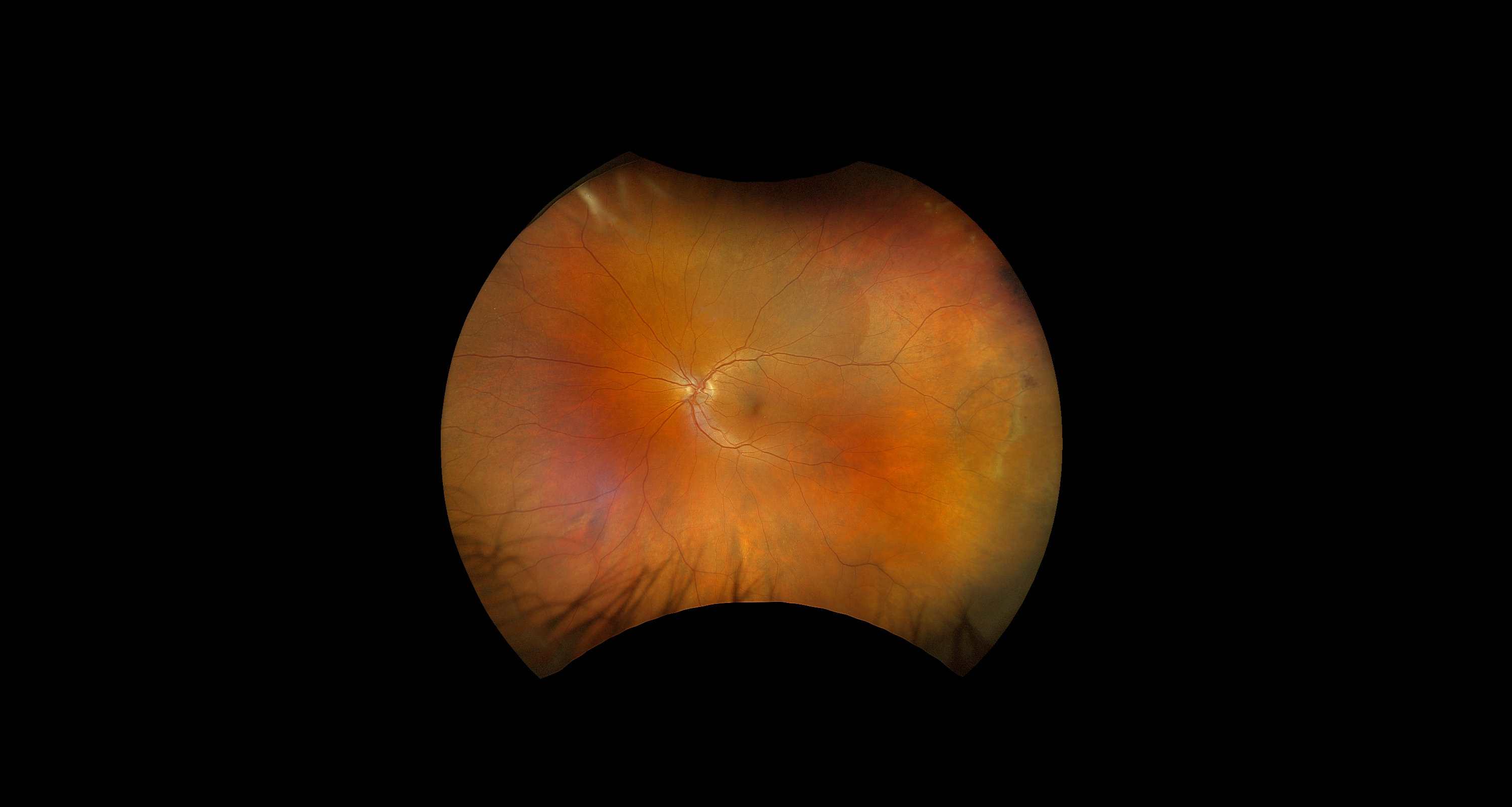
optomap multimodality captures high-resolution 200° ultra-widefield images that help detect, diagnose, and treat retinal disorders and diseases. This industry leading technology is being utilised by eye care professionals in practices around the world to enhance patient care and drive customer and staff retention. Advanced imaging technology allows clinicians to see more layers of the retina. This in turn allows optometrists and clinical practitioners to map, measure and assess the retina in closer detail with multimodality features.
An extensive body of clinical literature exists underlining the importance of evaluating as much of the retina as possible during an exam1. Studies demonstrate that imaging utilising ultra-widefield fundus capture with optomap guided SS-OCT can improve patient management and change treatment decisions. optomap gives a clearer image of the eye when compared to traditional fundus photography and works to support improved treatment of retinal diseases.
Common retinal diseases:
-Glaucoma
-Diabetes
-AMD
-Macular degeneration
-Geographic atrophy
Several2,3 large studies have underlined the importance of appropriately imaging the periphery to support the detection and management of disease in a variety of areas: telemedicine screening, diabetic retinopathy, age-related macular degeneration, vascular disease, paediatric retinal disease, inflammatory disease, and even some systemic diseases4.
Bringing the latest technological innovations into a practice or clinical setting not only supports ongoing treatment, but it can also help an optometry business drive revenue growth5,6. If a practice can become the trusted environment for a patient through the introduction of technology that improves patient care—the benefits to both an optometrist and patient can be seen for years7.
How Optos technology positively impacts a practice:
- Drive revenue and efficiencies with optomap 200◦ single image capture
- Develop patient loyalty through the embrace of eye care innovation
- Retain staff with training and utilisation of the latest technology
Value retinal imaging advancements in eye care
Introducing the latest retinal imaging technology into your practice helps improve patient relationships and increase the chance of early diagnosis8. An unintrusive optomap eye retinal examination can be completed within minutes and increases the speed of clinical care throughout a patient’s journey. The result is improved efficiency and an increase in revenue through the utilisation of advanced technology.
Consistently9 optomap UWF imaging has been demonstrated to capture the widest field of vision in a single capture of any imaging technology10. Capturing a larger field of view encompassing more of the retinal periphery is becoming increasingly important in a clinical setting. Given the clinical significance, it is vital a clinician understands the positive impact on the practice when obtaining those images.
Simon Browning, a leading optometry educator, believes strongly in the technological advancements and benefits Optos can bring to eye care:
“We need to change our approach on decision-making, address the challenges and perceptions around barriers, and implement positive change that will futureproof the industry. As a business owner, I’d encourage all optometrists to consider the benefits that come with ultra-widefield and hope one day to see an Optos device in every practice.”
Read Our eBook
[1] . Identification of diabetic retinopathy and Ungradable Image rate with Ultrawide field Imaging in a national Teleophthalmology program. Ophthalmology. 2016. [1]
[2] . Potential Efficiency Benefits of Nonmydriatic Ultrawide field Retinal Imaging in an Ocular Telehealth diabetic retinopathy program. Diabetes Care. 2013.
[3]. A novel hybrid fixed and mobile ultrawide field imaging program for diabetic tele-retinopathy screening. Ophthalmology Retina. 2019.
[4]. A novel hybrid fixed and mobile ultrawide field imaging program for diabetic tele-retinopathy screening. Ophthalmology Retina. 2019.
[5]. Comparison of Early Treatment Diabetic Retinopathy Study Standard7-Field Imaging With Ultrawide-Field Imaging for Determining Severity of Diabetic Retinopathy. JAMA Ophthalmology. 2018.
[6]. Peripheral Lesions Identified on Ultrawide Field Imaging Predict Increased Risk of Diabetic Retinopathy Progression over 4 Years. Ophthalmology 2015.
[7]. Paediatric retinal conditions imaged by ultra-wide field fluorescein angiography. Ophthalmic surgery, Lasers and imaging Retina. 2013.
[8]. Wide-field retinal imaging in the management of non-infectious posterior uveitis. American Journal of Ophthalmology, 2012.
[9]. Peripheral Findings and Retinal Vascular Leakage on Ultra-Widefield Fluorescein Angiography in Patients with Uveitis. Ophthalmology Retina. 2017.
[10]. Comparison of Wide-Field Fluorescein Angiography and Nine-Field Montage Angiography in Uveitis. American Journal of Ophthalmology. 2014.
[11]. Assessment of diabetic retinopathy using two ultra widefield fundus imaging systems the Clarus and Optos systems. Hirano. BMC Ophthalmology. 2018.
[12]. Zeiss 510(k) K181444.
[13]. Comparison of widefield confocal laser scanning ophthalmoscopy and broad line fundus imaging in retinal ophthalmoscopy and broad line fundus imaging in retinal disease in routine clinical practice. ARVO 2019.