Benefits of Multimodal Ultra-widefield Retinal Imaging
Optos offers multi-modal imaging with all of its ultra-widefield devices. Having both ultra-widefield and four images captured in less than one second has been shown to enhance pathology detection and disease management as well as improve practice and clinic flow. Ultra-widefield multi-modal imaging is important across all access points of patient care - screening, detection, diagnosis, and treatment.
Optos UWF multi-modality retinal imaging devices
Daytona, the first of the next generation of Optos devices was developed to facilitate comprehensive eye exams, document findings, and assist eyecare professionals with patient care. The technology delivers ultra-widefield images, in five modalities, and 200 degrees, from the central pole to the periphery, in less than ½ second.
MonacoPro combines optomap ultra-widefield technology with SD-OCT creating a 200°, single-capture retinal image and can display a six-image overview including color, AF, and OCT of both eyes in as little as 90 seconds.
Following MonacoPro, Optos introduced Silverstone, the only multi-modal platform that integrates UWF retinal imaging with integrated swept-source OCT, allowing optomap-guided OCT scanning across the retina and into the far periphery.
Most recently, a new imaging modality has been introduced, expanding the optomap UWF retinal imaging modalities available. In addition to optomap color (red/green), sensory Red-free, choroidal, autofluorescence (AF), fluorescein angiography (FAF) and indocyanine green angiography (ICGA) modalities, California is available with a new ultra-widefield color rgb (red/green/blue) image.
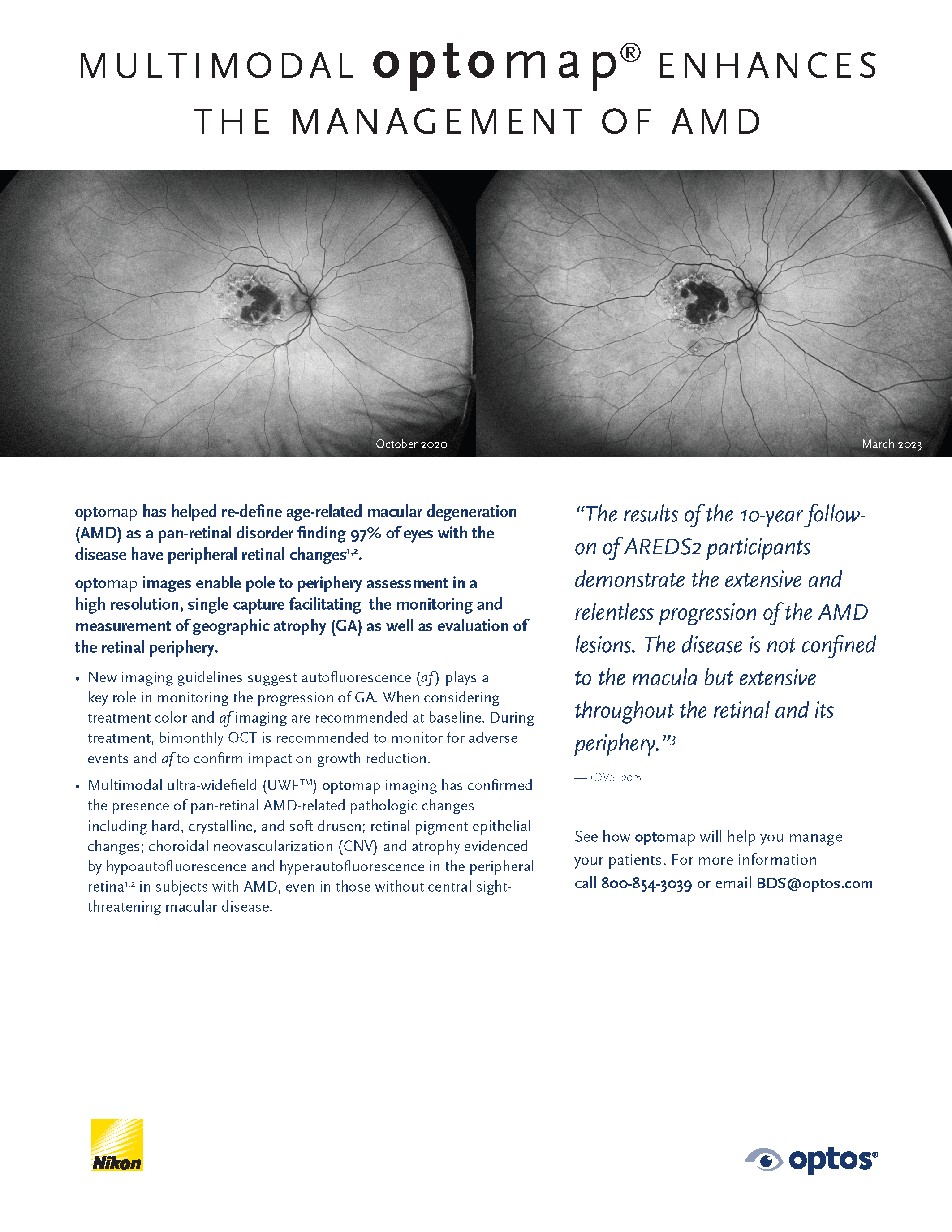
Multimodal optomap enhances the management of AMD
optomap has helped re-define age-related macular degeneration (AMD) as a pan-retinal disorder finding 97% of eyes with the disease have peripheral retinal changes. optomap images enable pole-to-periphery assessment in a high resolution, single capture facilitating the monitoring and measurement of geographic atrophy (GA) as well as evaluation of the retinal periphery.
New imaging guidelines suggest autofluorescence (af) plays a key role in monitoring the progression of GA. When considering treatment color and af imaging are recommended at baseline. During treatment, bimonthly OCT is recommended to monitor for adverse events and af to confirm the impact on growth reduction.
Read the whole clinical study here
If you're ready to put the power of muli-modality, ultra-widefield retinal imaging in your practice or clinical setting, contact us today.