Optos Clinical Papers and Summaries
This material is designed as a searchable reference resource to support clinical decision-making. The information contained here should be used as general guidance when viewing optomap and OCT images from Optos devices. The differential diagnosis should be made under the direction of the responsible physician. These images were taken on the latest ultra-widefield optomap devices.
Featured Clinical Summaries
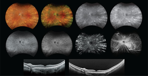
optomap Multimodality UWF Imaging Improves Clinical Practice
Multimodality UWF retinal imaging for streamlining capture & review to improve clinic flow & efficiency.
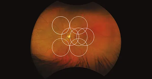
optomap-Assisted Ophthalmoscopy Improves Sensitivity of BIO by 30%
A study in Eye and Brain found good agreement between optomap-guided and traditional fundus examination.
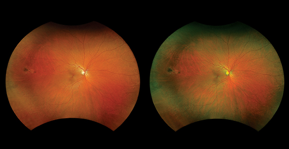
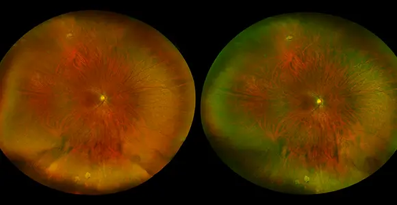
optomap color rgb - A More Natural UWF View
optomap color rgb - significantly superior in image diagnostic information compared to the gold standard.
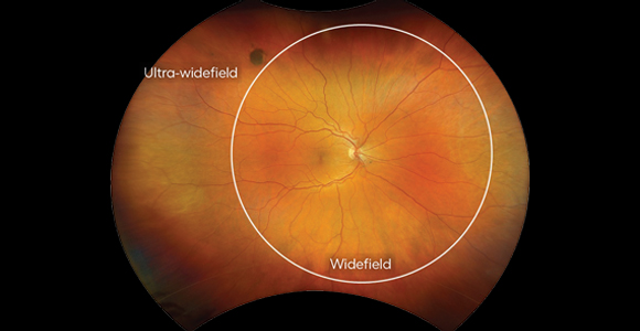
optomap Defining Ultra-widefield
optomap is the only single capture UWF retinal image, by definition.
optomap Improves Clinic Efficiency
optomap enables the reduction of patient wait times & improves clinic efficiency.
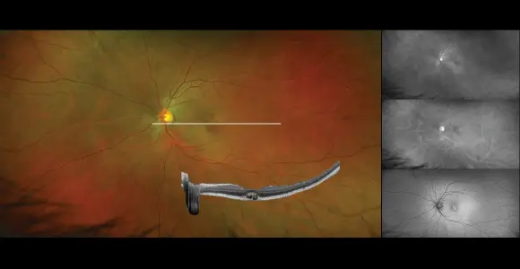
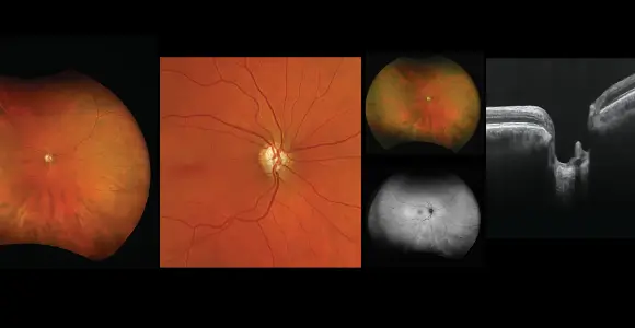
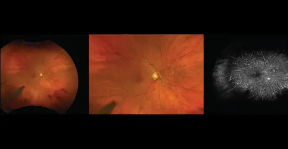
optomap-registered OCT Can Increase the Sensitivity & Efficiency of Exam
optomap with SD-OCT increases the identification of macular pathology over fundus imaging alone by 29.4%.
optomap Equivalent to ETDRS
Studies confirm the equivalence of optomap to ETDRS Gold Standard for grading diabetic retinopathy.
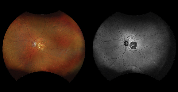
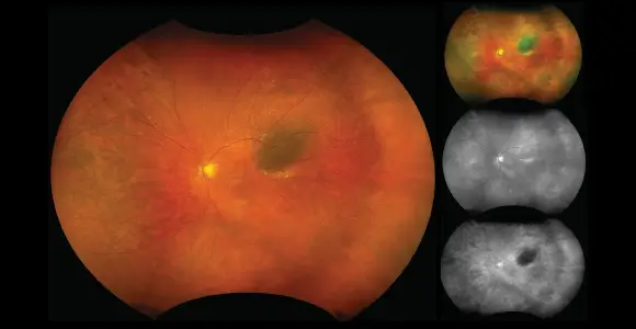
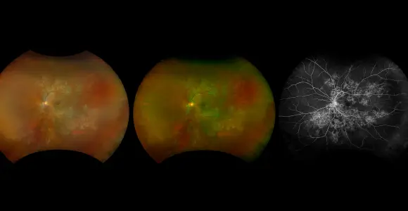
Multimodal optomap Images Enhance the Management of AMD
optomap has helped re-define AMD as a pan-retinal disorder.
optomap Equivalent for Glaucoma Assessment
Published clinical studies suggest that optomap may play an essential role in glaucoma management.
optomap has Excellent Agreement with Exam for Peripheral Lesions
optomap has a sensitivity of 89% for peripheral retinal lesions when compared to indirect ophthalmoscopy.
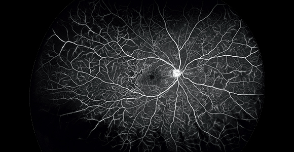
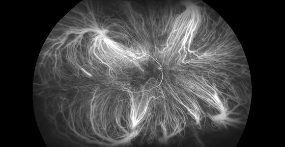
optomap fa Key Indicator to Predict the Progression of PDR
Research using optomap fa reveals that 50% of eyes with baseline PPL are at high risk for DR.
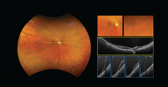
optomap-guided OCT Improves Patient Management
optomap-guided OCT imaging impacts clinical decision making in 84% of cases.
optomap Redefines Standard of Care for Inflammatory Disease
Patient management changed because of effective capture inflammatory and infectious disease.
67% of Eyes have Peripheral Findings on optomap icg
UWF icg reveals abnormalities in the peripheral retina that may be missed on conventional icga imaging.
optomap Improves Myopia Management
95% of HM optomap patients with drusen-like deposits in the peripheral retina have pathologic myopia.
optomap Strengthens Pre- and Post-Cataract Surgical Care
Multimodal optomap imaging supports the assessment of retinal health pre- and post- cataract surgery.
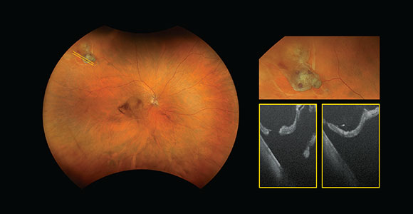
optomap is More Informative and Cost Effective for Ocular Oncology
optomap provides more information than traditional CFP for diagnosis & management of ocular oncology.
optomap for Pediatric Retinal Imaging
Studies suggest that optomap is an essential element to the screening & management of pediatric patients.
optomap for Telemedicine Programs
Ocular telemedicine programs that include optomap have a nearly double detection rate of DR.
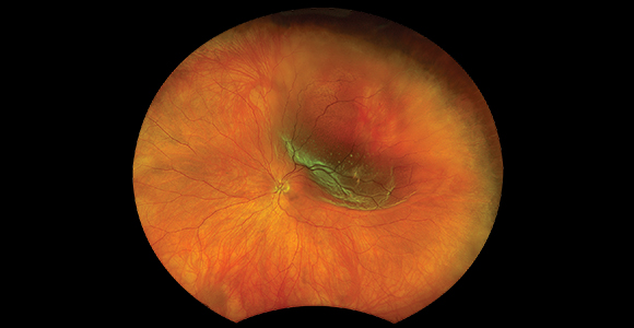
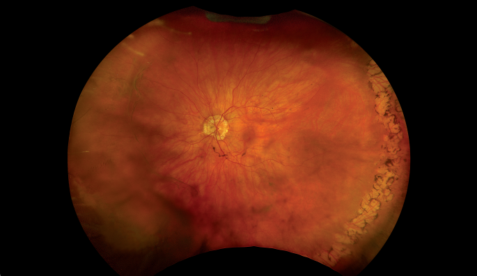
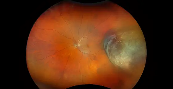
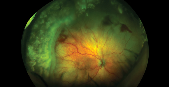
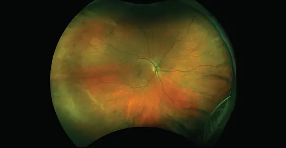
optomap for Retinal Detachments
optomap is equivalent to dilated fundus examination and is consistent with intraoperative findings when assessing retinal detachment.