Ultra-widefield Imaging in the Management of Diabetic Retinopathy
As the rates of diabetes in the US continue to rise, ultra-widefield (UWF™) retinal imaging technology from Optos, facilitates a more rapid visualization of a much larger area of the retinal periphery, resulting in earlier diagnosis, better evaluation, and more effective treatment options.
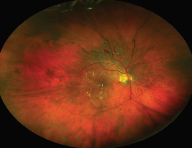
UWF color image of proliferative DR with DME
Improved Detection
retinal images are obtained more quickly without the need for dilation. In addition, UWF color imaging capable of viewing more than 80 percent or 200° of the retina allows for better viewing of the periphery for diagnostic purposes.
UWF imaging and UWF fluorescein angiography (FA) improves detection and classification by revealing early signs of diabetic retinopathy that may be missed in traditional assessment. The presence of peripheral lesions identified by UWF imaging may indicate a greater degree of diabetic retinopathy severity. Studies have also found that peripheral retinal ischemia, identified by UWF FA, is associated with diabetic macular edema. In patients with recurrent vitreous hemorrhage after diabetic vitrectomy, the presence of peripheral neovascularization, peripheral nonperfusion, and late peripheral vascular leakage was detected by UWF imaging at higher rates. These results indicate the need to assess peripheral retinal vessels by UWF FA after diabetic vitrectomy and implement additional treatment when necessary to prevent complications.
In patients with recalcitrant diabetic macular edema, larger areas of retinal nonperfusion identified by UWF FA were associated with a greater severity of diabetic retinopathy. These findings led to the development of an ischemic index which supports the conclusion that areas of untreated retinal nonperfusion may produce biochemical mediators of ischemia and disease progression. A recent study has shown that peripheral lesions identified with UWF imaging predict an increased risk of diabetic retinopathy progression over a four-year period, regardless of baseline disease severity. These results indicate the importance of visualizing the peripheral retina when evaluating the risk of diabetic retinopathy progression.
More Effective Management
Clinical evidence from studies incorporating UWF imaging is changing the understanding of diabetic retinopathy as well as the treatment approaches based on evolving clinical standards. A diagnosis of severe nonproliferative diabetic retinopathy based on findings from the peripheral retina is likely to be made earlier than one based on the criteria of the ETDRS 4-2-1 rule. In addition, even if only a few microaneurysms are identified in a standard assessment, an UWF FA may show extensive peripheral nonperfusion which indicates a greater degree of diabetic retinopathy severity. An earlier diagnosis means treatment can be initiated earlier, slowing disease progression.
Treatment approaches for diabetic retinopathy and diabetic macular edema are evolving as the findings of UWF imaging increase the understanding of these diseases. UWF images reveal that diabetic macular edema is actually a disorder of the peripheral retina which secondarily involves the macula, and it may present in either an acute or chronic phase.
In diabetic patients, retinal ischemia is caused by the shutting down of retinal capillaries due to the loss of pericytes under hyperglycemic conditions. Vascular endothelial growth factor (VEGF) is released in response to the ischemia. Studies have found that diabetic macular edema can be improved or resolved by treating the ischemic retina with a combination of VEGF inhibition and targeted panretinal photocoagulation (PRP). The examination of the retinal periphery with UWF imaging plays a key role in evaluating the efficacy of diabetic retinopathy treatment options, including PRP and VEGF inhibitor therapy.
Without UWF technology from Optos retinal imaging technology, pathology in the retinal periphery may go undetected, resulting in greater disease progression and possible loss of vision. In his article for Retina Today, Paul E. Tornambe, MD, suggests that the “standard of care must evolve to the point when every patient with diabetes undergoes periodic optomap® imaging.”
For more information on the clinical benefits of UWF imaging, please contact us directly at BDS@optos.com.