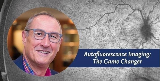
Ultra-widefield Autofluorescence Imaging: A Game Changer - Webinar Invitation
As an eyecare professional, providing comprehensive exams is tantamount to patient care. By adding tools, such as ultra-widefield (UWF™) retinal imaging with multiple modalities, the ability to detect pathology, which may be missed with single-image modality and/or conventional narrow-field fundus photography, is a game changer.
UWF retinal imaging is performed by a specially designed scanning laser ophthalmoscope (SLO) that generates a high-resolution digital image covering 200 degrees (or about 82 percent) of the retina, in a single capture. By comparison, conventional 7 standard field (7SF) ETDRS and fundus camera photographs produce a relatively narrow view (75 degrees or less) of the retina. The SLO simultaneously scans the retina using two low-power lasers (red – 633 nm and green – 532 nm) that enable high-resolution, color imaging of retinal substructures. The resulting UWF digital image – optomap or optomap af – UWF retinal imaging utilizing fundus autofluorescence (FAF), is produced.
What is Fundus Autofluorescence?
FAF, is an imaging modality used to provide information on the health and function of the retinal pigment epithelium (RPE). Over time, the retinal photoreceptors naturally age and produce a metabolic waste known as lipofuscin. Lipofuscin is the fatty substance found in the retinal pigment epithelium. Excessive amounts can be caused by the aging retina, certain retinal diseases and/or the progression of diseases.1 It has been thought that excessive levels of lipofuscin could affect essential RPE functions that contribute to the progression of age-related macular degeneration (AMD).2 These findings have also been shown to have prognostic value and help to predict which eyes are at greater risk of progression to advanced disease.3 Typically, FAF imaging has clinical applications in AMD, central serous retinopathy, choroidal tumors and nevi, inflammatory diseases, inherited disease, optic nerve head drusen, pattern dystrophies, retinal toxicity and retinal detachments. FAF excitation wavelength is between 480-510 nm, with an emission wavelength from 480-800 nm.4 optomap af uses a wavelength of 532nm to capture an image.
One of the main benefits of using the green wavelength, instead of blue, in FAF is its ability to see additional detail in the fovea, as the blue light tends to be absorbed by the high concentration of xanthophyll pigments. SriniVas Sadda, MD says this about FAF, “One advantage of longer-wavelength [green] light is that there is less absorption by the crystalline lens of the eye, which is quite autofluorescent with blue light, especially in patients with cataracts.”
FAF in Practice
Mr Simon Browning, has used optomap technology for nearly 20 years and was the first optometrist globally to use a Daytona. Since its installation, Simon has lectured at ARVO and the European Academy demonstrating how optomap af may be used to monitor regression of macular drusen in line with lifestyle and dietary changes. Simon believes ‘that optomap af is a game changer in optometry and that ‘optometrists are no longer required to fix a patients vision, but are there to preserve it.’4
Simon goes on to say, ‘Standard fundus imaging of the retina allows eyecare professionals to look at the structure of the retina, whereas optomap af imaging allows you to look at the functionality of the retina, often you will find that FAF imaging will show completely different images to the traditional colour image. Knowing this it allows eyecare professionals to react much faster to changes in the retina at a much earlier stage.’
Join Us for a Complimentary Webinar

optomap af imaging is a vital feature in monitoring and treating pathology in the retina, join us at 7pm AEDT (8am GMT) on Tuesday, 20th February for a free webinar hosted by Simon Browning to find out more about FAF imaging, identifying pathology using the FAF function as well as practice benefits.
Click here to register: https://Optos.is/ImagingEducation
1Holz, F. S.-V. (2010). Atlas of Fundus Autofluorescence Imaging. Heidelberg, Germany: Springer-Verlag.
2Delori, F. G. (2001). Age-Related Accumulation and Spatial Distribution of Lipofuscin in RPE of Normal Subjects. IVOS, 42(8), 1855-1866.
3Sadda, S. (October 2013). Evaluating Age-Related Macular Degeneration With Ultra-widefield Fundus autofluorescence. Retina Today.
4The Nuts and Bolts of Fundus Autofluorescence Imaging, 2017, American Academy of Ophthalmology.