Ultra-Widefield (UWF) Retinal Imaging Helps Manage Telehealth Evaluation of Diabetic Retinopathy (DR)
Earlier this year, we shared a report, which suggested UWF imaging’s potential to improve diagnostic efficiency for conditions such as DR and diabetic macular edema. In a separate study published in Diabetes Care, non-mydriatic UWF retinal imaging, in comparison to non-mydriatic fundus photography (NMFP), showed potential in improving “the efficiency of ocular telehealth programs evaluating DR and diabetic macular edema.
According to Healio, a total of 1,633 patients with diabetes were tested for DR and diabetic macular edema with NMFP for this study, and 2,170 patients were tested for the same conditions using UWF retinal imaging. There were no significant differences in age, duration of diabetes, gender, ethnicity or insulin existed among the two groups. Non-mydriatic UWF imaging was found to show a 71% reduction in the ungradable rate and a 28% reduction in image evaluation time, with an average image evaluation time of 9.2 minutes as opposed to 12.8 minutes for NMFP.
In addition, non-mydriatic UWF imaging was found to be 4.6% better at identifying cases of DR, as well as 2.6% better at identifying the most severe, vision-threatening cases of DR.

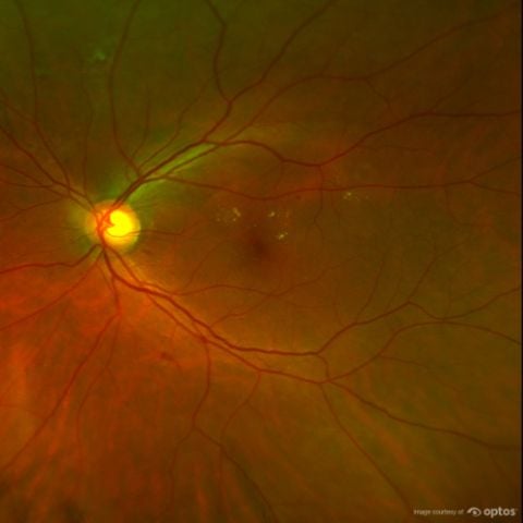
An optomap® image showing diabetic macular edema.
As diabetes is a leading cause of blindness in individuals between the ages of 20 and 74, it’s important to make sure these patients understand the risk their condition poses to their vision and that you not only encourage them to receive regular eye exams, but offer them innovative imaging technology to provide them with better quality care. Optos’ UWF retinal imaging technology and devices can help you see more by providing a 200-degree field of view of the retina. Contact us today to learn more about how our technology can benefit your patients, practice and diagnostic and disease monitoring capabilities.