Ultra-widefield Imaging is Changing Patient Management
In case studies, peer-reviewed papers, and a growing body of real-world practice, the ocular health community is improving patient management with the wider use of ultra-widefield (UWF™) imaging. Like all diagnostic breakthroughs, adoption of UWF imaging has been a long term process, paced by the accumulation of validated clinical experience. But now, more than a decade after UWF imaging was first introduced, the evidence is overwhelming that UWF imaging may have the potential to improve the diagnosis and management of a significant group of ocular diseases and conditions.
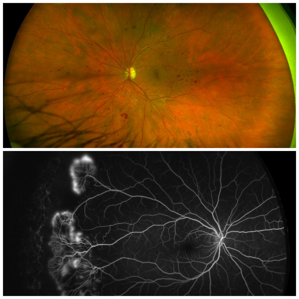
Top: Diabetic Retinopathy, California – Courtesy of Paulo Stanga, MD. Bottom: Retinopathy of Prematurity, Daytona, Proviewed
UWF imaging technology captures a 200-degree image of the retina – which enables ocular health practitioners to capture peripheral retinal images that can not be captured with conventional imaging methods. Starting with color (red and green) optomap imaging, Optos has systematically extended its UWF-based technology into a multi-modal platform that supports fundus autofluorescence (optomap af), fluorescein angiography (optomap fa) and indocyanine green angiography (optomap icg).
Where does UWF imaging have potential to improve diagnosis and treatment?
Proliferative Sickle Cell Retinopathy (PSR)
In a first of its kind case study1, researchers used multi-mode UWF imaging to examine the vascular changes associated with PSR, a complication of sickle cell disease that impacts the retinal periphery. The study, which covered a small number of cases, reported that the ability of UWF imaging to detect areas of non-perfusion and neovascularization missed by conventional imaging methods led to changes in treatment in some patients2. The authors also noted that UWF imaging provided added information that improved risk assessments of vision-loss from vitreous hemorrhage and tractional retinal detachment, two consequences of PSR progression.
Diabetic Retinopathy (DR)
Multiple studies using UWF imaging to diagnose and characterize DR have validated the technology’s value in multiple roles: as a screening tool; as a diagnostic technology that frequently detects more pathology than traditional methods; and as prognostic guide, via the detection of peripheral lesions that can help forecast the risk and severity of DR progression3.
Retinal Vasculitis
Studies report the diagnosis and treatment of retinal vasculitis is often altered when UWF imaging is used. In one report4, physicians changed patient management decisions in 51% of the non-infectious retinal vasculitis cases where optomap fa was added to the diagnostic work-up. A similar study by the same group in more mainstream, non-infectious uveitis found that management was also change 48% of the time5.
Retinopathy of Prematurity (ROP) and Pediatric Care
In the case of ROP and pediatric care in general, UWF imaging’s non-invasive characteristics and comparative ease-of-use are increasingly making it part of routine patient management. In treating ROP, one study5, reported Optos UWF imaging generated clinically useful post-treatment images while avoiding two disadvantages of traditional imaging methods – the need to bring the instrument in direct contact with the infant’s eyes and to anesthetize some infants to immobilize their eyes and/or limit head motion.
Imaging the Peripheral Retina Changes the Playing Field
As in any endeavor, the ability to visualize the entire playing field can change the game.
Doctors treating Central Serious Chorioretinopathy (CSC) in a small group of patients reported6 that their use of optomap icg images not only revealed more widespread disease, but also proved useful is identifying areas of choroidal hyperpermeability.
Practitioners also report UWF imaging contributing to the management and treatment of Age-related Macular Degeneration (AMD), retinal breaks and tears, Uveitis, Retinal Vein Occlusion (RVO)7, and retinal and choroidal vascular tumors8.
More clinical applications are being reported in conferences and papers, all reinforcing the premise that wider visualization of the retinal periphery can provide practitioners critical insights that can’t be made using conventional imaging.
***
Ocular disease frequently presents complex paths of progression, multiple therapeutic options, and varying paths to recovery. Ultra-widefield imaging can help manage your patients’ progress with unique, high-value diagnostic data that informs treatment plans and improves health outcomes.

Sources:
- How UWF Imaging is Improving the Management of Sickle Cell Retinopathy. September 23. https://blog.optos.com/how-uwf-imaging-is-improving-the-management-of-sickle-cell-retinopathy/
- Ibid, page 3
- Tornambe, Paul E., Ultra-Widefield Imaging: Advancing the Understanding and Management of Diabetic Retinopathy, Retina Today, April 2015
- Campbell JP, Leder HA, Sepah YJ, etal. Widefield retinal imaging in the management of noninfectious posterior uveitis. Am J Ophthalmol. 2012;154:908-11.
- Non-contact Ultra-widefield imaging of Retinopathy of Prematurity Using the Optos dual Wavelength scanning Laser Ophthalmoscope. Eye. 2013.
- Pang CE, Shah VP, Sarraf D, Freund KB, Ultra-Widefield Imaging with Autofluorescence and Indocyanine Green Angiography in Central Serous Chorioretinopathy, American Journal of Ophthalmology (2014), doi: 10.1016/j.ajo.2014.04.021
- Ultra-widefield Fundus Imaging: A Review of Clinical Applications and Future Trends; Nagiel, et al; Retina 2016
- Heimann H, Jmor F, Damato B. Imaging of retinal and choroidal vascular tumours. Eye. 2013;27(2):208-216. doi:10.1038/eye.2012.251.