optomap single-capture UWF Continues to Provide Clinical Value and Enhance Patient Care
This spring, the pandemic abruptly brought global apprehension and uncertainty. Medical practitioners desperately endeavored to navigate increased safety protocols while continuing to provide optimal care for their patients. Consequently, forecasters began to observe that reliance on medical technology solutions that can support safer exam scenarios would dramatically increase. During this challenging transition, Mitch Reinholt, OD discovered that one of his favorite diagnostic tools became more valuable than ever.
In April, Dr. Reinholt found himself with a bare-bones staff, providing some telemedical visits, but ultimately bringing patients in and doing most of the exams on his own. He found that his California helped to expedite exams and reduce patient exposure as well as time in the office. California, like all optomap UWF retinal imaging devices, capture over 200 degrees of the retina in 1/2 second. It provides a dynamic image that can be enlarged and manipulated to see into the retinal layers and with autofluorescence to assess function, as well. Reinholt explained that he could capture the image quickly, then withdraw to review it and evaluate if further diagnostics were required.
Reinholt acquired his California device in 2019 primarily for clinical value and the ability to get unmatched retinal information, but also because the ease and speed of acquisition expedited the exam process and impressed his patients. Additionally, he found that the technology further streamlines the exam process by enabling him to quickly image children and other difficult to examine populations.
In the short time that he has had the technology, it quickly established its clinical value by capturing several pathologies that he feels might have been otherwise undetected such as far peripheral retinal holes, lattice, and white without pressure. He also recounts an instance when he imaged a long-time patient and discovered a large, peripheral operculated hole that had gone undiscovered previously because the patient had always refused to be dilated.
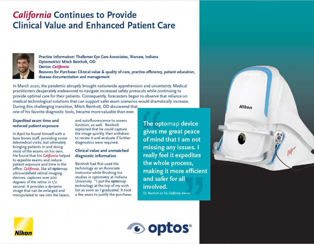
As we begin to embrace a new normal, Dr. Reinholt contemplates the uncertainties of the future and remains adamant that optomap technology will be essential to the function of his practice. Doctors are concerned with the number of patients in the office at a time, and in minimizing the amount of time each patient has to be there. “I definitely plan on imaging everyone. This way I can review the information thoroughly prior to examining the patient,” says Dr. Reinholt. He adds that the dynamic and comprehensive image gives him incomparable amounts of information to help direct the rest of the exam. He notes that prior to the pandemic he had an 83% acceptance rate for optomap screening exams and that his patients were always happy to see their image. Going forward he plans that he will image every patient and that now, more than ever, they appreciate limiting extended close exposure in the traditional exam. “The optomap device gives me great peace of mind that I am not missing any issues. I really feel it expedites the whole process, making it more efficient and safer for all involved.”
Optos helps doctors to discover and document the retina with little or no face-to-face interaction and optomap single-capture UWF imaging has been to shown to increase practice flow and patient engagement. Go here to read Dr. Reinholt’s full testimonial. Visit our website today to put the power, efficiency, and safety of optomap in your practice.