The OPS Scientific Exhibit Image Competition
The Ophthalmic Photographers’ Society (OPS) holds an image competition annually at the American Academy of Ophthalmology (AAO). The competition is open to all members and always features a number of interesting images that represent the best in retinal imaging across a number of different categories.
The judge’s panel for the competition is made up of OPS members and ophthalmologists who select the images that best represent the categories of the competition, which range from conditions like retinal angiography to images demonstrating surgical techniques, and even creative submissions under “Eyes as Art.” The winning images are then put on display at the OPS Scientific Exhibit on the AAO Exhibit Floor during the show.
However, prior to the Exhibit’s opening, a third set of judges reviews the images one last time and selects what OPS describes as “the most outstanding image from the show.” That image is honored with the Csaba L. Martonyi Award, which is named for Csaba L. Martonyi, CRA, FOPS, a longtime member of the OPS who encouraged professional imagers to put their “effort and skill into producing images that serve a medical purpose and demonstrate technical and artistic perfection.”
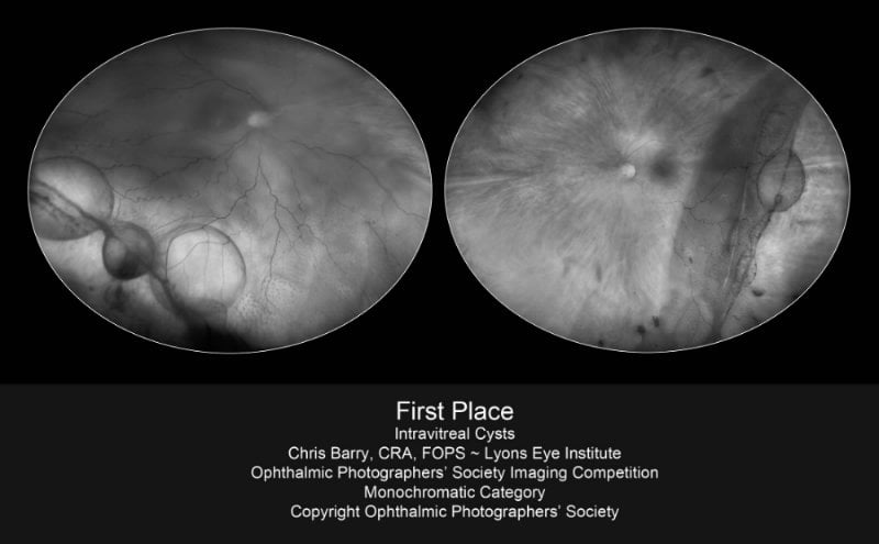
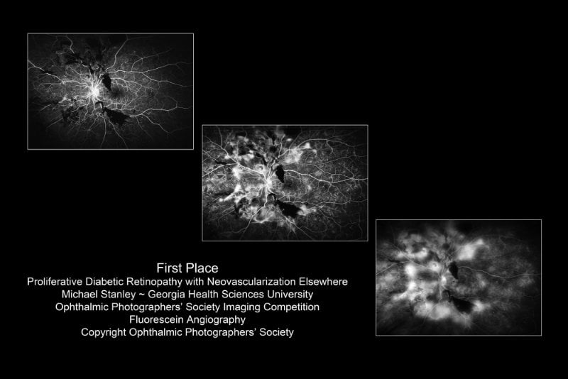
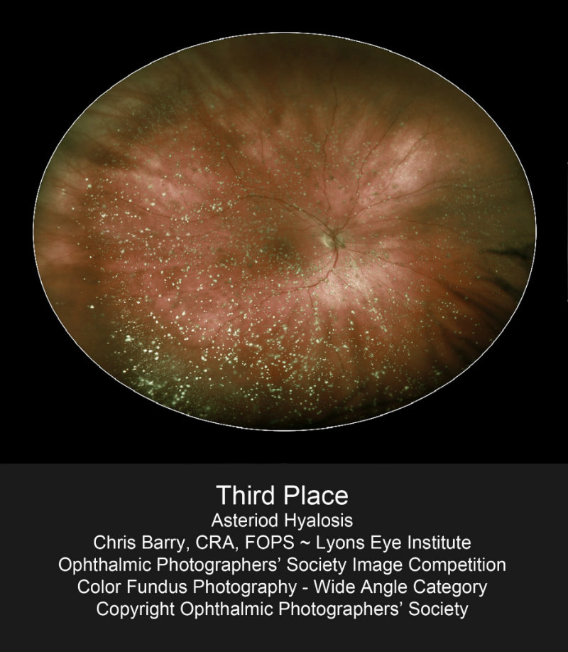
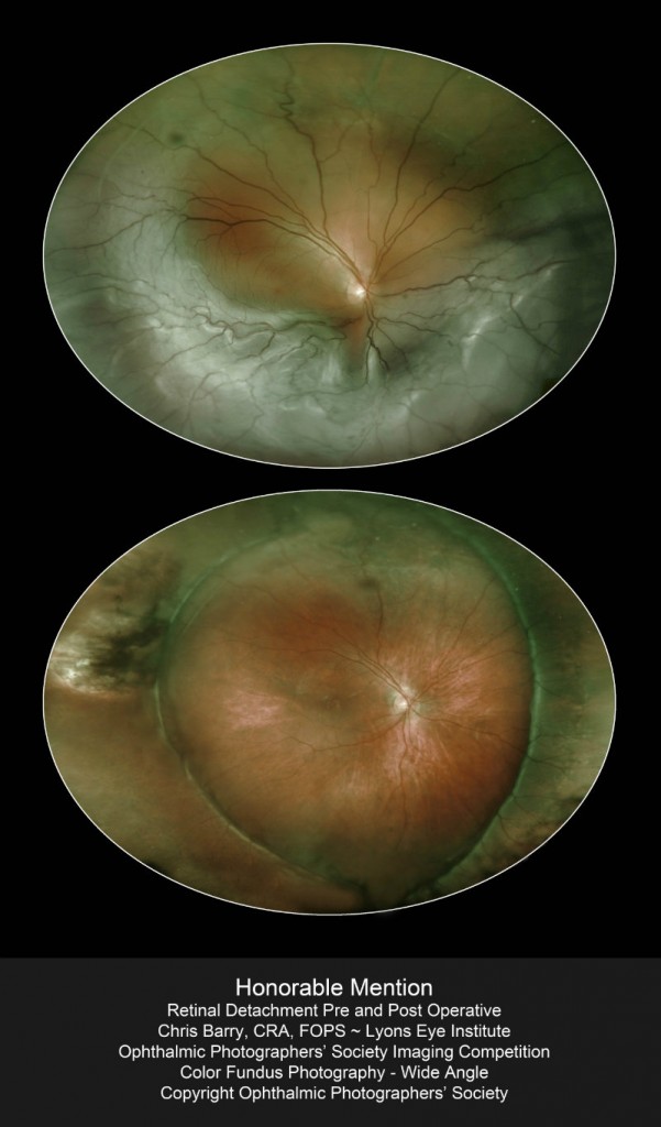
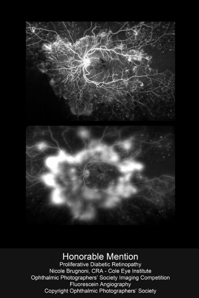
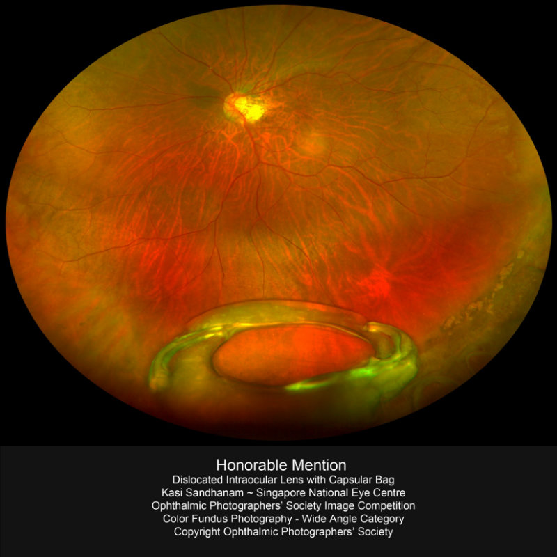
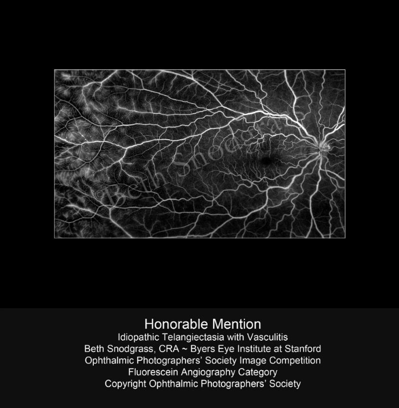
In the 2012 competition, Optos was honored to have eight optomap images selected as winners in the Fundus Photography Wide Angle Category, Monochromatic Photography Category, and Fluorescein Angiography Category. In total, Optos images received two First Place Awards, one Third Place Award, and five Honorable Mentions. Below is a look at some of our winning images (Please click on the images for a larger view).
To view more of our images, please visit the Optos website. These images offer significant clinical value that any practitioner can benefit from viewing.
Are you a practitioner interested in learning more about Optos’ ultra-widefield retinal imaging devices and the many benefits they offer to you, your practice, and your patients? Request a consultation with an optomap professional today.