See the Full Picture with Ultra-Widefield Retinal Imaging
When you’re driving to a place you’ve never visited before, a map is a helpful tool that can aid you finding the place you need to be. A street view provides you with an up-close look at where you’re going, but it may cut off certain signs or landmarks you’ll need to look for. A wider view of a map shows you more of what you need to see in order to arrive at your destination with ease.
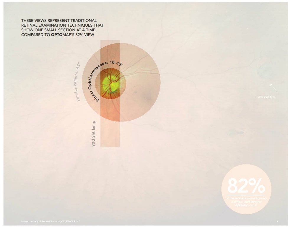
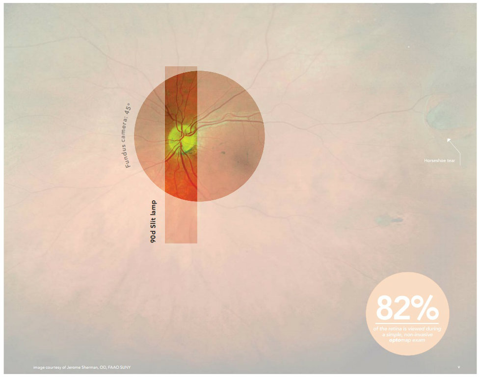
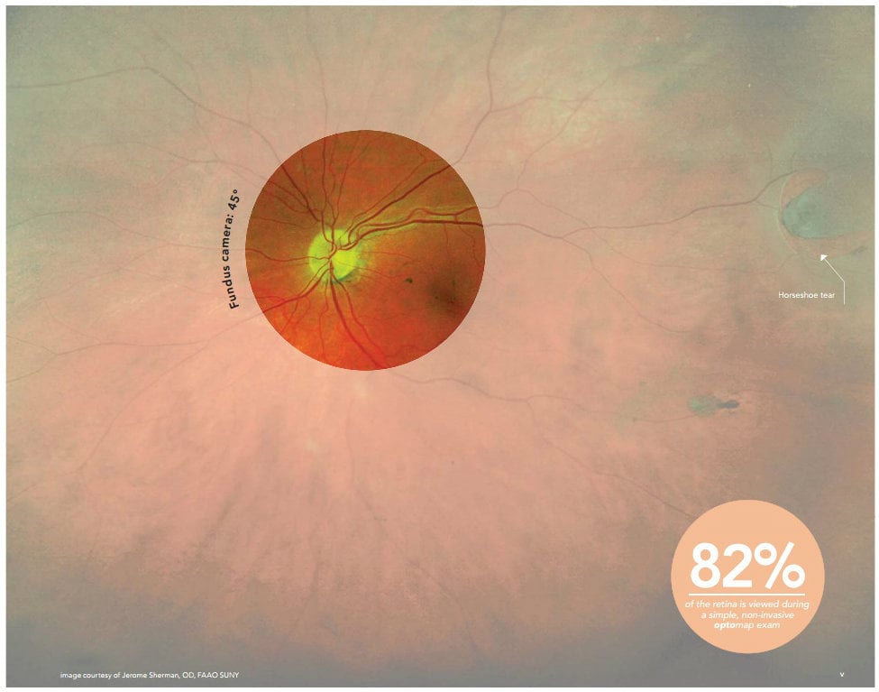
Similar things can be said for an eye exam. In a standard eye exam, conventional equipment limits you to a 45-degree field of view of the retina, showing very small portions of the retina at a time. With such limitations, there is a good possibility that signs or symptoms of an eye or systemic disease are left out of your field of view.
Ultra-widefield retinal imagining provides you with a more complete picture of your patient’s eyes. Optos’ ultra-widefield technology allows you to see up to 200 degrees of the retina in just one capture. With a wider field of view, you have a better opportunity to detect issues like retinal detachment or tears you might have otherwise missed.
The images below compare images captured through traditional techniques with an image captured with Optos’ optomap exam. Take note of the difference in perspective.
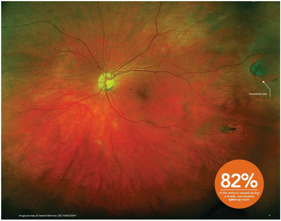
The difference is clear – you aren’t seeing the full picture if you aren’t using ultra-widefield retinal imaging. To learn more about the advantages Optos’ ultra-widefield technology can offer to you, your practice, and your patients, please visit our website or contact us for more information.