Earlier Detection of Alzheimer’s May be Possible Through Eye Exams
Memory loss and confusion are just a few of the symptoms associated with Alzheimer’s disease. And sadly, the disease is already quite advanced by the time patients begin exhibiting symptoms and an official diagnosis is given, making it rather difficult to treat. Past studies have indicated that there is potential for eye exams to aid in earlier detection of Alzheimer’s disease, as have advances in research Healio Optometry recently reported on, which have revealed there may be potential for a new method of earlier detection of Alzheimer’s, perhaps long before patients start presenting symptoms.
Neurologists have determined the presence of amyloid beta protein deposits as a “biomarker” of Alzheimer’s. Amyloid can also accumulate in the eye, leading researchers to theorize, “If a correlation can be made between the amyloid in the eye and the amyloid in the brain, then it would be possible to diagnose [Alzheimer’s] by looking into the eye.”
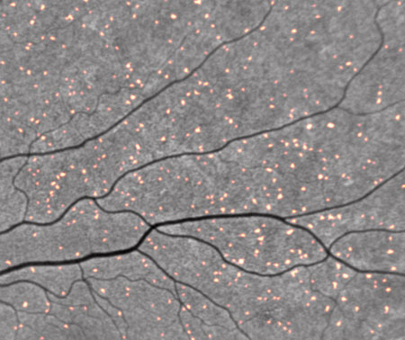
With this theory in mind, researchers are working to develop tests that will detect these amyloid beta deposits in the eyes. One test, currently referred to as the “Retinal Amyloid Index,” takes a scan similarly to conventional retinal imaging devices, after the patient has taken a curcumin compound orally, to detect the deposits. The curcumin “crosses the blood-brain and blood-retina barrier and binds with high affinity to amyloid beta plaque – particularly amyloid beta 42, which is the most toxic” and naturally creates a fluorescent signal that can be captured during a retinal scan.
Keith L. Black, MD, shared that fluorescence detectable by retinal imaging would be “directly proportional to the amount of amyloid protein in the back of the eye, which correlates with the amount of amyloid protein in the brain.” Because amyloid beta proteins begin to accumulate long before the obvious signs of Alzheimer’s, this test could potentially allow for diagnosis decades earlier than before.

Amyloid protein in the eye detected with the Retinal Amyloid Index.
Source: NeuroVision via Healio.com
The other test, called “Sapphire II,” works to detect the same amyloid deposits in the crystalline lens by way of a “fluorescent ligand marker applied topically in an ointment” the evening before the procedure. The procedure consists of taking fluorescent measurements with a device that provides a numerical score directly related with the detected level of amyloid.
Carl Sadowsky, MD, shared that the process is much like an amyloid PET scan of the brain. “We measure pathology; it’s not diagnostic of [Alzheimer’s]. It’s an adjunct that you add to your clinical consideration. So if a patient has amyloid in the brain, and they have clinical symptoms of AD, it makes us more confident of the diagnosis.”
While each study on this topic may have a different outline, they all have the same objective of being able to diagnose Alzheimer’s earlier. We’re excited to see how eye exams may lead to better diagnosis and treatment of Alzheimer’s in the coming years as more research is conducted on the topic. As we’ve shared before, many systemic diseases, such as chronic myelogenous leukemia, can be detected through Optos’ optomap ultra-widefield retinal imaging exam.
Contact us today to learn more about how our technology can be an aid in detecting and diagnosing eye and health issues your patients may not even know they have.