November is Diabetic Eye Disease Awareness Month
Diabetic eye disease is a leading cause of blindness and vision loss,1 and, according to the World Health Organization, the incidence of diabetes worldwide is escalating.2 It is estimated that by the year 2035, the number of people worldwide with diabetes will have soared to almost 600 million.3 The National Eye Institute (NEI) reports that there are another 86 million American adults who have pre-diabetes.
Diabetic eye disease describes a group of eye conditions that include diabetic retinopathy (DR), glaucoma, diabetic macular edema and cataracts. DR is often reported as the most common form of diabetic eye disease. It is a serious complication of diabetes mellitus (DM), afflicting one third of all people with the disease, and it is the leading cause of blindness among the working population in the world.4
In its report, “Diabetic Eye Disease Projected to Increase Among U.S. Population,” the NEI states that there are currently 7.7 million people ages 40 and older who have DR, and this number is expected to increase to around 11 million by the year 2030. With advances in technology and medicine, adequate management and regular eye examinations, sight loss associated with diabetes may be prevented in 98 percent of cases.5
Primary interventions in the prevention of sight loss in people with diabetes include regular, effective screening programs to detect diabetic eye disease earlier combined with education to encourage patients to undergo yearly eye examinations. Many clinicians agree that UWF is an important part of these examinations, this was recently highlighted in an article by Dr. Paul E Tornambe where he calls for the integration of ultra-widefield (UWF™) imaging as both a practical and clinical asset to the management of patients with diabetes.6 Given the statistics mentioned above, there is potentially a significant challenge for eye care facilities to provide effective and efficient protocols for screening for diabetic eye disease. UWF continues to evolve to address specific patient requirements, including looking at non-mydriatic imaging alternatives which are also designed to be more time efficient.7 optomap® is clinically proven and its multimodal capabilities place it as a leader in imaging patients with diabetes to support the detection of the diabetic retinopathy.
While technology has been advancing, patients continue to go blind. There are still reports that patients fail to attend their annual eye examinations.8 One of the reasons cited for non-attendance is that some patients are not fully aware of the connection between diabetes and eye disease, often believing that if their eyes are asymptomatic, they need not attend an eye examination.9 Integrating UWF imaging could support an improvement in patient compliance and engagement, educating patients about peripheral findings that they may not detect, but which may need urgent attention if left unchecked.
Diabetic patients may have to submit to multiple instillations of mydriatic drops to achieve full pupil dilation, leaving some patients with discomfort due to light sensitivity. Routinely screening patients using non-mydriatic, UWF imaging technology helps ease this anxiety and may reduce the need for dilation at every visit. The speed, multimodal functionalities, patient-centric considerations and practical benefits have established the optomap as a proven clinical technology in the fight against vision loss. In less than half a second, optomap is able to capture a 200 degree image (up to 82 percent) of the retina through an undilated pupil. From a patient education perspective, the resulting images may serve to provide the patient with a visual representation of the link between their physical well-being and their ocular health. Clinically, UWF has a proven record as valuable technology in a variety of screening programs. It is the ease with which optomap images are captured,10 annotated, stored and shared that facilitate optimal practice efficiency and resource use.
As this potential diabetic eye disease epidemic looms, there is a clear need to work towards the development of a cohesive partnership between patients and eye doctors. Patient engagement, cooperation, and comfort are key factors to ensuring that diabetic eye disease is detected and treated as early as possible, and optomap imaging technology fully supports this collaboration. The incorporation of ultra-widefield technology to support eye screening protocols and routine workflows has the potential to alleviate the projected increased burden of diabetic eye disease on clinical resources, and it also supports improved patient education, compliance and sight loss prevention in at-risk individuals.
Learn more about the clinical benefits of utilizing ultra-widefield optomap in your practice or clinic.
References
- https://www.nei.nih.gov/sites/default/files/nehep-pdfs/GM_DED_drop-in%20article_2014.pdf
- https://www.who.int/diabetes/global-report/WHD2016_Diabetes_Infographic_v2.pdf?ua=1
- James Kang Hao Goh, BSc, Carol Y Cheung, PhD, Shaun Sebastian Sim, MBBS, Pok Chien Tan, MBChB, Gavin Siew Wei Tan, MBBS and Tien Yin Wong, MD, PhD – Retinal Imaging Techniques for Diabetic Retinopathy Screening. Journal of Diabetes Science and Technology 2016, vol 10(2) 282-294 – DOI:10.1177/1932296816629491 dst.sagepub.com
- Yau JWY, Rogers SL, Kawasaki R, et al. Global Prevalence and Major Risk Factors of Diabetic Retinopathy. Diabetes Care. 2012;35(3):556-564. DOI: 10.2337/dc11-1909.
- Nentwich MM, Ulbig MW. Diabetic retinopathy – ocular complications of diabetes mellitus. World Journal of Diabetes. 2015;6(3):489-499. DOI: 10.4239/wjd.v6.i3.489.
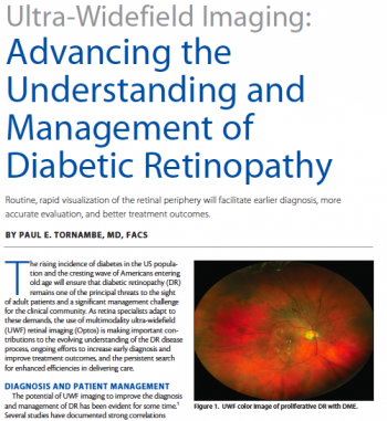
- Paul E. Tornambe, MD, FACS. Cover Story – Ultra-Widefield Imaging: Advancing the Understanding and Management of Diabetic Retinopathy. Retina Today, April 2015 https://retinatoday.com/2015/04/ultra-widefield-imaging-advancing-the-understanding-and-management-of-diabetic-retinopathy
- Liu SL, Mahon LW, Klar NS, et al. A randomised trial of non-mydriatic ultra-wide field retinal imaging versus usual care to screen for diabetic eye disease: rationale and protocol for the Clearsight trial. BMJ Open 2017;7:e015382. DOI: 10.1136/bmjopen-2016-015382.
- Fisher MD, Rajput Y, Gu T, et al. Evaluating Adherence to Dilated Eye Examination Recommendations Among Patients with Diabetes, Combined with Patient and Provider Perspectives. American Health & Drug Benefits. 2016;9(7):385-393.
- M J E Huber, S A Smith, S E Smith. Mydriatic drugs for diabetic patients. British Journal of Ophthalmology, 1985, 69, 425-427.
- David M Brown. Advancing the Detection and Management of Diabetic Retinopathy with Ultra-widefield Retinal Imaging. US Ophthalmic Review, 2017;10(1):23-6. DOI: https://www.touchophthalmology.com/articles/advancing-detection-and-management-diabetic-retinopathy-ultra-widefield-retinal-imaging