3D Wrap™ Allows Practitioners to Give Patients a “Tour” of Their Eye
Ask any doctor who has been practicing for many years, and they’ll probably agree that educating patients on a particular diagnosis is one of the best things you can do to help them understand what’s going on with their body and to improve adherence to treatment plans or ongoing visits. At Optos, we not only believe in providing practitioners with the resources to educate their patients, but also to show a patient exactly what they see in an retina exam. One of those tools is our innovative and interactive 3D Wrap™ tool.
– The patient’s iris color from capture settings
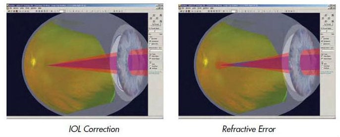
– Refractive errors range from -10D to +5D, including the ability to demonstrate myopia and hyperopia through simulation of the shape of the model eye
– Demonstrating how myopia and hyperopia affect the patient’s vision by displaying different ray types, simulating near or distant objects, as well as an overlap of the two
– IOL selections such as Monofocal and Multifocal lenses
This is just a glimpse at how Optos’ 3D Wrap™ can let practitioners better educate their patients on vision issues, the inner workings of their eyes and more. Those interested in learning more can contact us today to speak with a representative. If you currently use 3D Wrap™, leave a comment below to share your experience with your fellow practitioners.
It is important to note that 3D Wrap™ is not for diagnostic purposes.