How UWF Imaging is Helping Correlate Retinal Pathology and Systemic Disease
Ultra-widefield retinal imaging (UWF™) is helping to identify retinal pathology that could have prognostic value in estimating the risk of some systematic diseases.
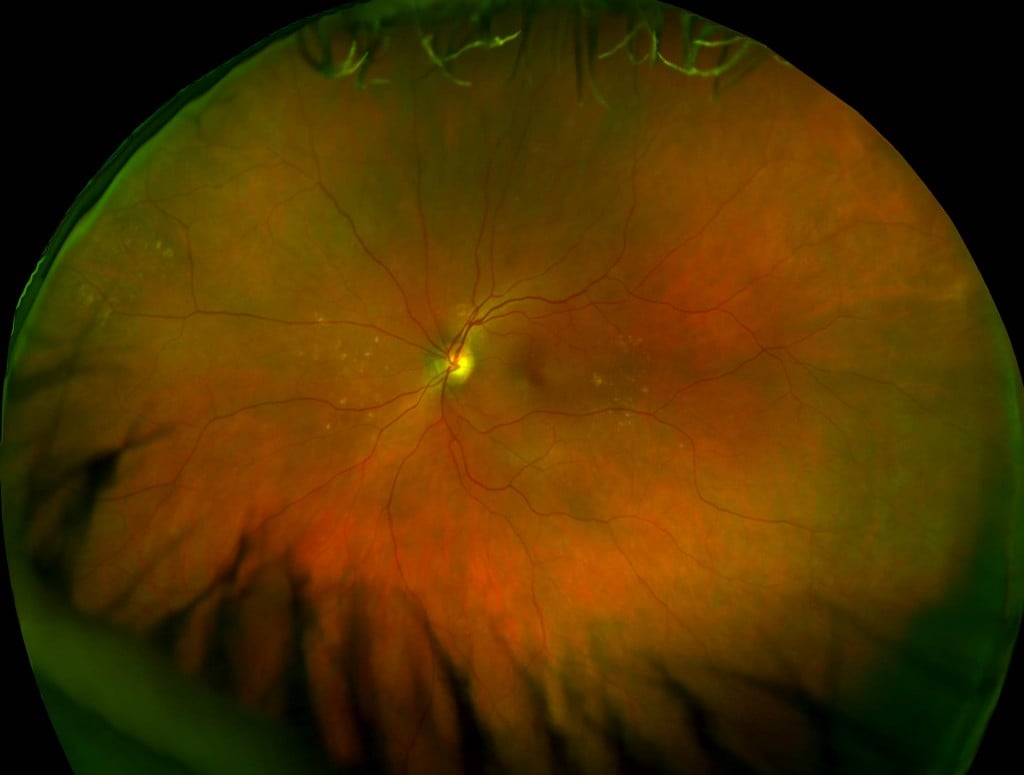
Color optomap image, peripheral drusen, which according to research, may be related to detecting the onset or progression of alzheimer’s disease (AD)
A topic of continuing interest in the health community is the use of retinal pathology to predict the risk of systematic diseases such as Alzheimer’s. If retinal pathology with significant prognostic value can be identified and this pathology can be quickly and reliably characterized, there’s the prospect for better and more cost-effective health screening on both an individual and community level. Multi-modal ultra-widefield imaging is beginning to be applied to research efforts that are exploring these correlations in greater detail.
About UWF Retinal Imaging
relatively narrow view (75 degrees or less) of the center-portion of the retina.
The SLO simultaneously scans the retina using two low-power lasers (red – 633 nm and green – 532 nm) that enable high-resolution, color imaging of retinal substructures. The resulting UWF digital image – optomap – is produced in a single capture.
Along with UWF color imaging, the technology supports UWF fluorescein angiography (FA), UWF fundus autofluorescence (FAF), red reflectance imaging (RR), and UWF indocyanine green angiography (ICG).
The Impact of Ultra-widefield Imaging and Alzheimer’s
Alzheimer’s disease (AD) is of growing impact on aging populations and to date there is no definitive, pre-mortem diagnosis for the disease. A variety of studies are using UWF imaging to characterize drusen formation, vascular health, and nerve fiber layer thickness in the peripheral retina and relate this to the onset or progression of AD.
— One early study4 compared a group of patients with AD and a control group. It found that optomap retinal images of the AD patients showed more peripheral drusen than the control group.
— In another study5 involving 60 patients with mild to moderate AD, the increasing presence of peripheral drusen correlated to worsened mental state.
— In a separate two year pilot study6 of 14 AD patients and 15 controls, all over 70 years of age, peripheral drusen progressed at a significantly higher rate in the AD patients versus controls.
— Decreased retinal blood flow may be another characteristic of AD. One study7 found that retinal blood flow was somewhat impaired in subjects with mild cognitive impairment and more impaired in subjects with AD. Retinal veins were found to be significantly narrower (135 microns in diameter) in subjects with AD versus the control group (158 um), suggesting a possible means to screen for decreased retinal blood flow and, potentially, AD.
— Early results from a comprehensive study8 that will characterize both peripheral drusen formation and NFL (nerve fiber layer) thickness in AD patients found that reduced NFL thickness and increased numbers of drusen were associated with increasing cognitive impairment and AD.
This and other similar research has yet to present definitive findings that presence and extent of peripheral drusen, reduced retinal blood flow, and NFL thinning can predict the onset of AD, but continuing studies have the potential in the not-distant future to make the widefield characterization of these pathologies a part of AD screening and diagnosis.
Similar work is underway in characterizing retinal vascular health in relation to other conditions. One of the requirements for this work is developing accurate algorithms that can be used to automatically process UWF images and provide critical data on blood vessel width. Studies to develop this technology are now being presented to the ocular health community.
The biggest challenges in understanding the prognostic value of retinal pathology are time and numbers. Multi-year and even multi-decade studies surveying large populations will be required in order to make statistically valid conclusions and change standards of clinical practice. UWF retinal imaging could well play a key role in this process by (1) enabling more complete characterizations of retinal health and (2) giving researchers and practitioners a proven, rapid and widely-available diagnostic tool on which to collect their base data and test their findings.

- Kawasaki R, Wong TY. Retinal vascular imaging for cardiovascular risk prediction. In Yogesan K, et al. (eds). Digital Teleretinal Screening. Springer-Verlag Berlin Heidelberg. 2012. 77-89.
- Klein R, Klein BE, Meuer SM. Retinal emboli and cardiovascular disease: the Beaver Dam Eye Study. Trans Am Ophthalmol Soc. 2003;101:173-182.
- Hanff, Thomas C., Sharrett , A. Richey, Mosley ,Thomas H., et al, Retinal Microvascular Abnormalities Predict Progression of Brain Microvascular Disease: An ARIC MRI Study, Stroke, 2014 April ; 45(4): 1012–1017. doi:10.1161/STROKEAHA.113.004166.
- https://optos.com/de-CH/Wir-uber-uns/Medienarbeit/Press-Room/Press-Releases/2011/Study-indicates-the-potential-value-in-the-detection-of-Alzheimers-Disease/
- Beguiristain MG, Zapata MA , Distefano LN, et al. Study of funduscopic and tomographic findings in the retina of patients with Alzheimer disease. Paper presented at: Association for Research in Vision and Ophthalmology Annual Meeting, May 3-7, 2015, Denver, CO.
- Aslam, Asma, Peto, Tunde, Barzegar-Befroei , Neda, et al, Assessing Peripheral Retinal Drusen Progression in Alzheimer’s Dementia: A Pilot Study Using Ultra-Wide Field Imaging, April 2014, https://iovs.arvojournals.org/article.aspx?articleid=2272166
- https://www.reviewofophthalmology.com/content/d/technology_update/i/1628/c/29944/
- https://www.alz.co.uk/sites/default/files/conf2015/OC001-Eleonora-Lad-Retinal-Imaging-Biomarkers-For-Early-Detection-Of-Alzheimers-Disease.pdf