UWF Case Study: Angiomatous Proliferation in ROP
History
A 15 year-old African American female with a history of bilateral threshold retinopathy of prematurity was seen for a routine annual visit. As an infant, she was treated with scatter laser photocoagulation two months after her birth. At this visit, she was asymptomatic with a visual acuity of 20/25 in the right eye and 20/15 in the left eye.
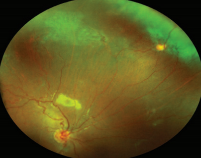
Color fundus photograph of the right eye showing a yellowish orange round lesion at 2 o’clock of the superonasal periphery.
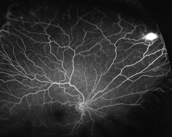
Mid-phase fluorescein angiogram of the right eye with focal, round area of hyperfluorescence in the superonasal periphery, approximately one disk diameter in size.
Examination
optomap® color images were obtained and showed a reddish orange, slightly elevated lesion at 2 o’clock in the far periphery of the right eye, anterior to the equator. Images also confirmed areas of previous laser treatment in the temporal periphery. optomap fluorescein angiogram images were obtained and showed the lesion to be hyperfluorescent and that the lesion had feeding and draining retinal vessels. The left eye did not have similar lesions, but did have evidence of previous laser treatment in the temporal periphery.
Discussion
optomap color and fluorescein angiography were used to document the appearance and investigate the characteristics of the peripheral lesion. The lesion was determined to be reactive angiomatous proliferation – a rare vascular proliferation which has been reported to occur in various chronic retinal diseases, such as retinitis pigmentosa and Coats’ disease. Most recently, it has been reported to have occurred in a 14 year-old female with a remote history of retinopathy of prematurity, similar to our patient. She underwent cryotherapy to the lesion, and early involution of the lesion was present one month following treatment.
Conclusion
optomap color and fa widefield imaging successfully captured, documented, and allowed the investigation of this rare vasoproliferative condition.
To obtain a copy of this case study, written by Anna Gabrielian, MD and Mathew W. MacCumber, MD, PhD, contact us at Optos.