Advanced Technology and Instrumentation Aids in Presentation of Eye Pathologies
As we’ve shared before, advanced technology and instrumentation are huge assets to practitioners because they not only help ensure a patient’s eyes are healthy, but also because such advanced equipment can detect potentially serious vision and medical conditions in their earliest stages. In doing so, a patient’s risk of going blind or becoming extremely ill is averted because treatment options can be presented much earlier on.
Review of Optometric Business recently shared three case studies in which Ed Liu, OD, of Foothills Optometric Group in Pleasanton, California, shared how an OCT scan and retinal imaging through an optomap® helped detect signs of macular degeneration, retinal detachment and leukemia.
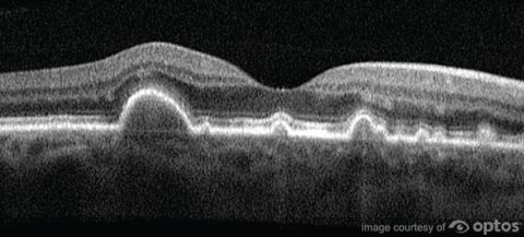
In the first case, Dr. Liu was unable to achieve 20/20 vision with the patient. This led him to complete an OCT scan, which allowed him to look at the patient’s optic nerve and retina. With this scan, Dr. Liu was able to detect drusen and other early signs of macular degeneration on a microscopic level that other devices would not be able to detect. Cases like this have paved the way for his five-doctor practice to use the OCT on a daily basis, allowing them to spot pathologies very early and create better patient outcomes.

Caption: Drusen detected through an Optos OCT/SLO scan.
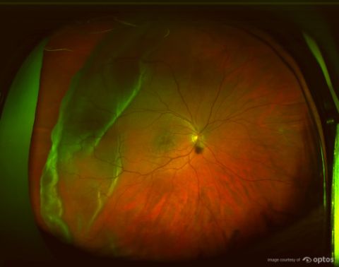
In the second case, Dr. Liu tells of a patient that came into the practice late one afternoon with complaints of floaters. The patient requested to make an appointment for a later time, but a staff member could tell there was a potential problem and encouraged the patient to be seen that day. A retinal scan was taken moments later, revealing a retinal detachment. Dr. Liu’s staff was able to email the images of the retinal scan to the local retinal specialist, and the patient was in surgery to have the detachment repaired just over an hour after the retinal scan was taken.

Caption: Retinal detachment as shown in an optomap® retinal image.
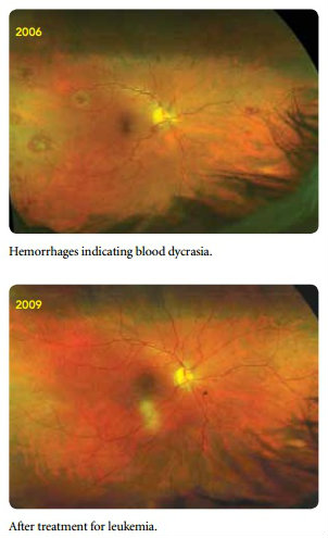
The final case Dr. Liu presented is that of a patient who was in for a routine optomap exam. The patient had no symptoms of any underlying issues, but the retinal scan revealed several hemorrhages in the retina. Dr. Liu referred the patient to the retinal specialist, providing images of the retinal scan, and about a week later the patient returned to inform Dr. Liu that the retinal scan helped lead to a diagnosis of leukemia. Because it was detected early enough, the patient was successfully treated and a follow-up optomap six months later showed the hemorrhages had cleared.

Caption: The top optomap image shows hemorrhages that indicated a case of chronic myelogenous leukemia (CML). The bottom image, taken about 3 years later, shows the effectiveness of the patient’s treatment for CML.
Are you interested in learning more about how advanced technology and instrumentation can better equip you to present eye pathologies to your patients? Visit the Optos website to read case studies in which our technology was able to help practitioners do just that. If you are interested in learning more about adding one of our OCT or Ultra-widefield imaging devices to your practice, please contact us.