Ultra-widefield Fundus Imaging: Clinical Applications and Future Developments
Over the past decade Optos has expanded the capability of its core ultra-widefield fundus imaging technology, and with that has come a widening number of clinical applications.
Ultra-widefield (UWF™) imaging technology – enabling the capture of a 200-degree view of the retina without dilation – gives ocular health practitioners imagery and diagnostic information that can’t be provided by conventional imaging methods. Starting with color (red and green) optomap imaging, Optos has systematically extended its UWF-based technology into a multi-modal platform that supports fundus autofluorescence (optomap af), fluorescein angiography (optomap fa) and indocyanine green angiography (optomap icg).
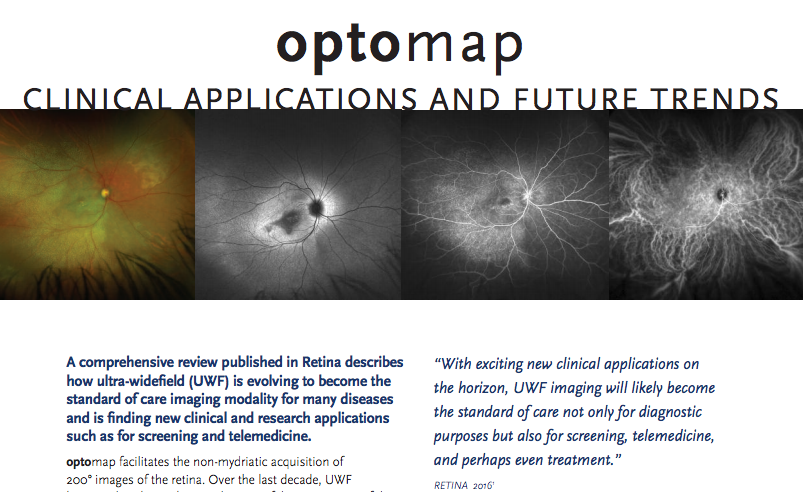
optomap – A comprehensive review published in Retina describes how ultra-widefield (UWF) is evolving to become the standard of care imaging modality for many diseases and is finding new clinical and research applications such as for screening and telemedicine.
Optos has also been incorporating the latest developments in image processing, providing users with important diagnostic and treatment management tools. In its latest software release, Optos has incorporated an advanced, proven stereographic projection algorithm that corrects for peripheral image variations that occur when a spherical image is flattened.
Clinical Applications
While the most common use of UWF technology may be in the diagnosis and treatment of diabetic retinopathy (DR), optomap imaging is also being used for characterizing pediatric retinal disease; age-related macular degeneration (AMD); retinal breaks and tears; uveitis, ocular oncology; central serous chorioretinopathy (CSCR); retinal vein occlusion (RVO); and a growing list of other diseases and conditions1. Practitioners have also reported that UWF imaging undertaken in an office setting can be used to document retinal pathology in infants, a procedure that might otherwise require anesthesia2,3.
Most studies comparing UWF with traditional photographs have determined the sensitivity and specificity to be similar but with additional peripheral information provided by UWF imaging1.
Some of the common benefits UWF imaging brings to practitioners include:
— The ability to document and evaluate evidence of disease in the central pole and retinal periphery in one image and
— Multi-mode capability, allowing pathology to be correlated with different diagnostic techniques.
Practitioners have reported that the richer, peripheral diagnostic data provided by UWF imaging can lead to changes in treatment management plans4. In particular, the availability of peripheral imagery is giving new insights into how ocular disease progresses and the impact of treatment.
The ease by which optomap imaging can be performed is also supporting its use in telemedicine programs, in which optomap images taken by a medical technician can be evaluated off-line by an ocular evaluation professional5.
Future Developments
Increased use of UWF imaging in clinical applications will be supported by four likely future developments:
— Pilot studies6 that used UWF imaging to characterize healthy eyes will expand into large-scale projects that will profile men, women, children, seniors, and other populations of interest. This will set the stage for more comprehensive diagnostic protocols and wider use of UWF imaging in both ocular health screening and treatment.
— Studies suggest that some retinal pathology visible on UWF images – for example, peripheral drusen, abnormalities in blood vessel width, and retinal emboli
– May be associated with a higher risk of Alzheimer’s disease7 and vascular diseases like arteriosclerosis, hypertension, myocardial infarction and stroke8. While much work is needed to validate these connections, it suggests a future role for UWF imaging as one of several low-cost, non-invasive diagnostic tools used to identify at-risk individuals.
— Continuing improvements in image processing software will enable practitioners to more accurately assess retinal pathology and better guide diagnosis and treatment. This includes the ability to process and measure blood vessel width and tortuosity9.
Deeper understanding of the healthy eye and retinal pathology have the potential to further widen the application of today’s UWF imaging technology. Combined with new advances in image processing and more diagnostic modalities, the clinical applications of ultra-widefield imaging will continue to grow.
For additional information on how UWF can fit into your eyecare environment, please contact us.
Sources:
- Ultra-widefield Fundus Imaging: A Review of Clinical Applications and Future Trends; Nagiel, et al; Retina 2016
- Non-contact Ultra-widefield imaging of Retinopathy of Prematurity Using the Optos dual Wavelength scanning Laser Ophthalmoscope. Eye. 2013.
- Non-contact ultra-widefield retinal imaging and fundus fluorescein angiography of an infant with incontinentia pigmenti without sedation in an ophthalmic office setting. Journal of AAPOs. 2013.
- Ultra-widefield Fundus Imaging: A Review of Clinical Applications and Future Trends; Nagiel, et al; Retina 2016
- Ultra-widefield Fundus Imaging: A Review of Clinical Applications and Future Trends; Nagiel, et al; Retina 2016
- Sadda S, Sagong M, van Hemert J, et al. Ultra-widefield imaging of the peripheral retinal vasculature in normal subjects, Ophthalmology, 2016 May;123(5):1053-9. doi: 10.1016/j.ophtha.2016.01.022. Epub 2016 Feb 17.
- Beguiristain MG, Zapata MA , Distefano LN, et al. Study of funduscopic and tomographic findings in the retina of patients with Alzheimer disease. Paper presented at: Association for Research in Vision and Ophthalmology Annual Meeting, May 3-7, 2015, Denver, CO.
- Pellegrini, Enrico et al. “Blood Vessel Segmentation and Width Estimation in Ultra-Wide Field Scanning Laser Ophthalmoscopy.” Biomedical Optics Express 5.12 (2014): 4329–4337. PMC. Web. 19 Apr. 2016.
- Pellegrini, Enrico et al. “Blood Vessel Segmentation and Width Estimation in Ultra-Wide Field Scanning Laser Ophthalmoscopy.” Biomedical Optics Express 5.12 (2014): 4329–4337. PMC. Web. 19 Apr. 2016.