How UWF Imaging is Improving Diagnostic Capabilities
Most advances in medical technology are very beneficial for improving a doctor’s abilities to both diagnose and treat a particular disease or condition. For ophthalmologists, advanced imaging devices are a must. But, as Ravi D. Patel, MD, an associate vitreoretinal surgeon and director of clinical research at Retinal Vitreal Consultants Ltd., Chicago, recently shared with Ophthalmology Management, there are few imaging devices that “change the way we think about a disease or fundamentally alter clinical management.”
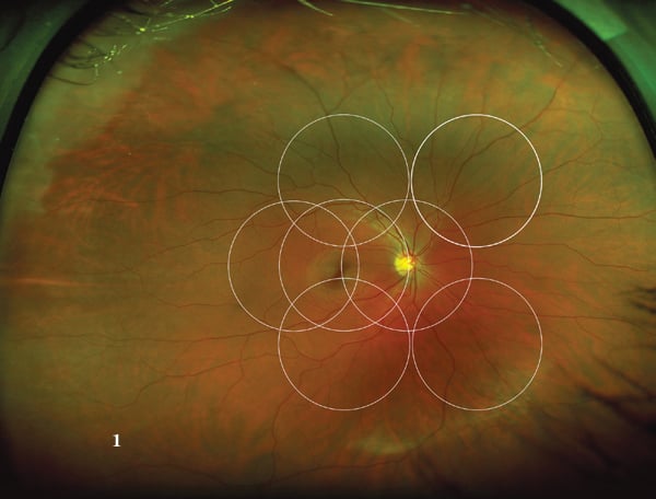
Dr. Patel focuses on ultra-widefield imaging (UWF), noting that this form of imaging has helped practitioners study areas they previously couldn’t with conventional imaging devices, especially in the vitreoretinal subspecialty. He looked at several different UWF imaging devices, but lists the ultra-wide views of up to 200° that Optos’ 200Tx scanning laser ophthalmoscope provides as incomparable to most. The enhanced view of the periphery, Dr. Patel shares, has helped lead researchers to some new clinical findings over the past few years, as well as new insights to the role of peripheral pathology in retinal vascular and degenerative diseases, among others. Dr. Patel also noted UWF imaging has helped uncover potential new disease markers.
Other benefits of newer UWF imaging devices Dr. Patel listed, from medical and practical standpoints, include:
- The ability to capture images simultaneously
- The ability to simultaneously detect several foci of neovascular dye leakage, as well as intraretinal comparisons of their severity
- Rapid image capture rates
- Ease of use
- Digital image storage
Overall, Dr. Patel feels that based on new research conducted using UWF imaging devices such as Optos’ 200Tx, technology is definitely changing the way ophthalmologists think about retinal disease and will provide improvement for diagnosis and patient management.

Caption: This image shows the 200° field of view captured through the Optos 200Tx scanning laser ophthalmoscope with the seven standard ETDRS fields superimposed, while also comparing the field of view with a 75° view.
Are you interested in learning more about all the benefits Optos 200Tx has to offer? Visit our website to learn more. There you’ll find information on all of our retinal imaging devices, as well as OCT imaging devices and diagnostic instruments. To speak with a representative, please request a consultation by contacting us.