Can my Optos Device diagnose Retinal Detachments?
The simple answer is “yes”, your Optos device can assist you in diagnosing the three different types of retinal detachment listed below.
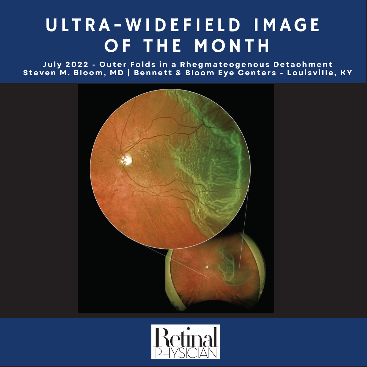
- Rhegmatogenous [reg-ma-TAH-jenous] – A tear or break in the retina causes it to separate from the retinal pigment epithelium (RPE), the pigmented cell layer that nourishes the retina, and fill with fluid. These types of retinal detachments are the most common.
- Tractional – In this type of detachment, scar tissue on the retina's surface contracts and causes it to separate from the RPE. This type of detachment is less common.
- Exudative – Frequently caused by retinal diseases, including inflammatory disorders and injury/trauma to the eye. In this type, fluid leaks into the area underneath the retina (subretina).
As a matter of fact, Optos itself was founded when a five-year-old boy was blinded after a regular eye exam failed to spot a retinal detachment, his father made it his life’s work to help eye care professionals by revolutionizing retinal imaging.
Over the past thirty years, Optos ultra-widefield retinal imaging technology has been used to image hundreds of millions of individuals in order to assist in the detection of retinal detachment, and other vision and life-threatening diseases. One of the most recent cases that was presented to highlight the technology was of outer retinal folds in a Rhegmatogenous retinal detachment, provided by Steven M. Bloom, MD of Bennett & Bloom Eye Centers.
Visit our website to find an eyecare professional near you utilizing optomap ultra-widefield retinal imaging technology.