Only two short years ago, the Ebola outbreak occurred in West Africa. Today, survivors are presenting with symptoms of post-Ebola Syndrome (PES) which include joint and muscle pain, and psychiatric, neurological, and eye problems1. Researchers from the University of Liverpool’s Institute of Translational Medicine have recently conducted a study of these survivors to determine what effects Ebola had on the retina.
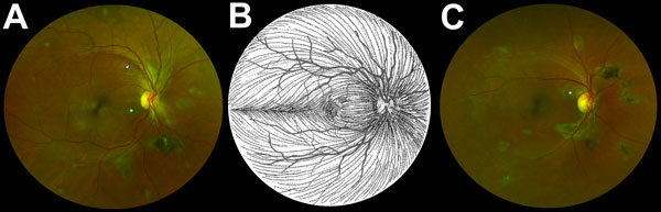
Composite scanning laser ophthalmoscope retinal images showing type 6 Ebola peripapillary or peripheral lesions, observed following the anatomic distribution of the ganglion cell axon (retinal nerve fiber layer), in a case–control study of ocular signs in Ebola virus disease survivors, Sierra Leone, 2016. A) Example 1, right eye. B) Illustration of the ganglion cell axon anatomic distribution. Courtesy of W.L.M. Alward. C) Example 2, right eye. Asterisks indicate curvilinear lesions distinct from the retinal vasculature. White arrowhead indicates retinal nerve fiber wedge defect.
The ocular research team was led by Paul Steptoe, MD and the research group compared the eye exams of 82 survivors who had previously reported ocular symptoms and a control group of 105 unaffected individuals. The Daytona from Optos was used to conduct the non-mydriatic ultra-widefield retinal imaging portion of the study. The results of this research which has been published in the Emerging Infectious Diseases Journal, shows that approximately 15% of Ebola survivors examined do have a retinal scar which appears specific to the disease2. According to researchers this is a reasonable conclusion based on the fact that the control group did not present with similar lesions and only demonstrated the common retinal issues that are present in a population prior to Ebola exposure.
Key Facts and Findings:
- 82 Ebola virus survivors (161 eyes, 2 missing retinal images, 1 prosthetic eye) and 105 never infected controls (208 eyes, 2 missing retinal images)
- Only type 6 subcategory retinal lesion was observed exclusively in the survivor group (14.6%) while 0% of the control group. In 50% of the survivors the lesion was bilateral.
- The Ebola retinal lesions did not appear to affect visual acuity.
“According to Dr. Steptoe: “The distribution of these retinal scars or lesions provides the first observational evidence that the virus enters the eye via the optic nerve to reach the retina in a similar way to West Nile Virus. Luckily, the scars appear to spare the central part of the eye so vision is preserved. Follow up studies are ongoing to assess for any potential recurrence of Ebola eye disease. Our study also provides preliminary evidence that in survivors with cataracts causing reduced vision but without evident active eye inflammation (uveitis), aqueous fluid analysis does not contain Ebola virus therefore enabling access to cataract surgery for survivors.”
Dr. Steptoe is a clinical research fellow at the University of Liverpool and a specialist ophthalmology trainee in the Mersey region, UK. His research interests include tropical ophthalmology with an emphasis on ophthalmic infections and uveitis. To find out more about the broad-ranging symptoms of Post Ebola Syndrome (PES), a clinical research team led by Dr. Janet Scott and Dr. Calum Semple assessed survivors in collaboration with staff at the 34th Regiment Military Hospital in Freetown, Sierra Leone.
Sources:
1,2 ‘Novel Retinal Lesion in Ebola Survivors, Sierra Leone, 2016’, DOI: 10.3201/eid2307.161608 & https://wwwnc.cdc.gov/eid/article/23/7/16-1608_article