optomap® Recognizing Pathology
This material is designed as a searchable reference resource to support clinical decision-making. The information contained here should be used as general guidance when viewing optomap and OCT images from Optos devices. The differential diagnosis should be made under the direction of the responsible physician. These images were taken on the latest ultra-widefield optomap devices.
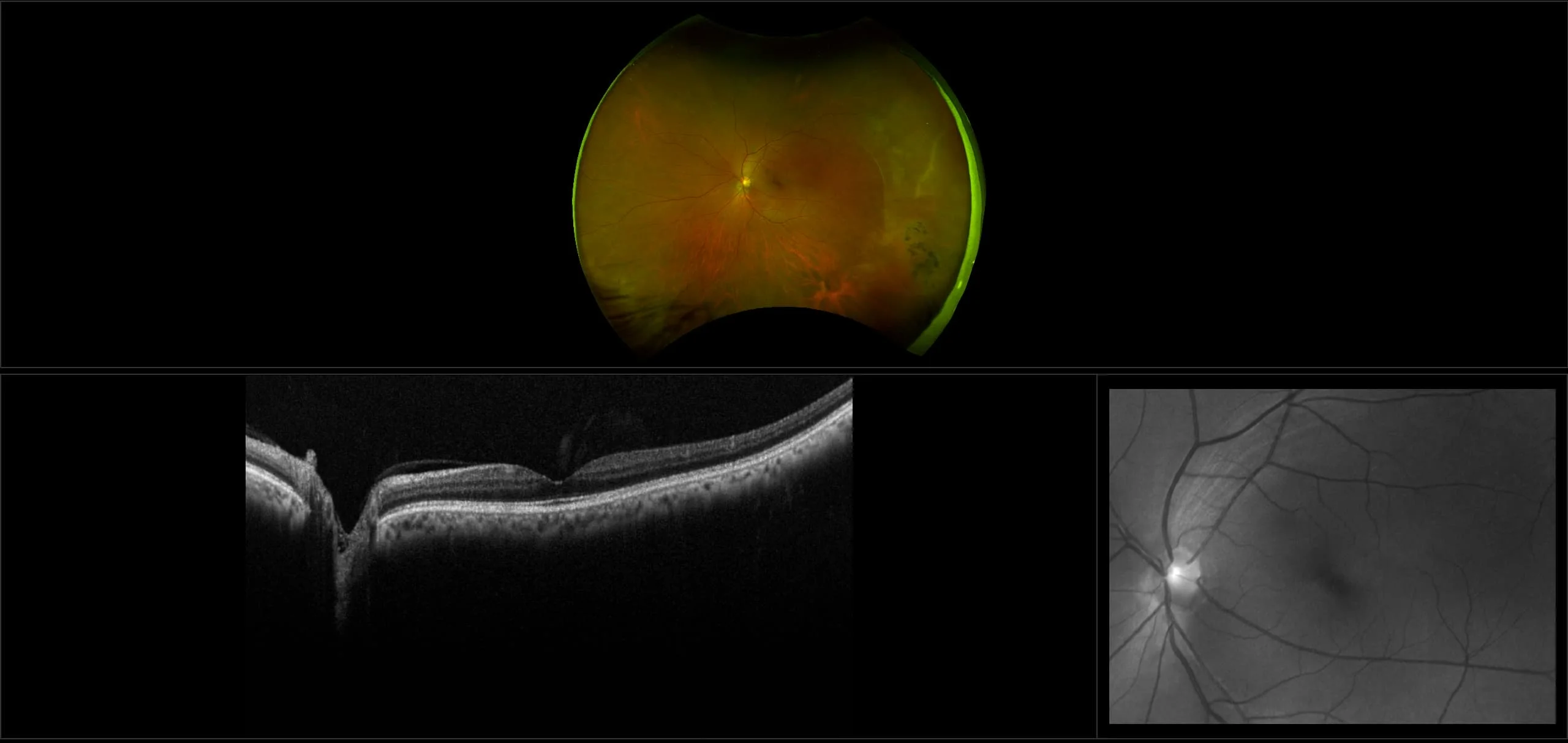
Retinal Pigment Epithelium (RPE) Hyperplasia
RPE hyperplasia results in a proliferation of RPE cells and thus, forms a pigmented retinal lesion which is very irregular in shape. All that is necessary is a proper stimulus to the RPE and this may be provided by physical stimulation such as vitreous traction or inflammation from infection or trauma. Sometimes a small area of this condition is called pigment clumping (gathering of RPE cells).