The ability to capture and document retinal detachment can prove indispensable when treating your patients. Not only can it help you determine the underlying cause of the detachment, it can prove a valuable teaching tool for your patients so they understand what is wrong and how their participation in their treatment will provide a better outcome.
To get a better understanding of this ailment and communicating properly to your patient, be sure to read through the case study from Optos below:
Source: Optos
History
A 60 year-old male visited the Retina Institute of Hawaii complaining of poor central vision in the right eye. He began to notice vision loss inferiorly one week prior, which progressed to central vision loss two days prior to exam. The patient was CF with eccentric viewing.
Examination
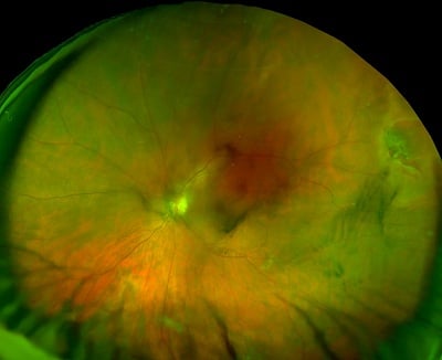
Upon a dilated fundus examination, there was a macula-off superior Rhegmatogenous Retinal Detachment with a superotemporal horse-shoe tear. optomap plus images were captured to document the retinal detachment and horseshoe tear and assist in explaining the treatment plan to the patient. A pneumatic retinopexy was immediately performed which reattached the macula.
The patient laid in the office in a face-down position for 30 minutes and was instructed to maintain the face-down position overnight. On the second day, his vision was 20/400 and the optomap revealed that the macula was reattached, and much of the subretinal fluid shifted inferiorly. It was likely he alternated between the face-down and head-up position. Laser retinopexy was performed to seal the horseshoe tear found in the superior temporal quadrant (one day after pneumatic). The patient was then asked to maintain head tilted to left, so that the pneumatic retinopexy would continue to close the horseshoe tear. Patient returned three days later with most of the subretinal fluid reabsorbed, macula attached, and the horseshoe tear sealed. His visual acuity measured 20/200 at this point.
Discussion
In this case, optomap plus images demonstrated rapid and accurate documentation and assisted in proper diagnosis and effective treatment. More importantly, it aided in explaining the disease and treatment strategy to the patient. Without optomap plus images, it would have been more difficult to explain the steps of treatment to the patient. This likely increased patient compliance and prevented the patient from having to go to the operating room to achieve an excellent outcome.
Conclusion
The patient returned home and was instructed to keep his head tilted to the left. He followed up four days later with the subretinal fluid completely reabsorbed. His visual acuity measured 20/40 with a nuclear sclerotic cataract. The horseshoe tear was well treated with laser retinopexy
Contact Optos to learn how to integrate optomap’s positive effects of early detection and treatment for your patients’ best outcomes.