The statistics are clear. Over the next decade optometric and ophthalmic practices in North America, Europe and throughout the developed world will identify and treat an increasing number of patients with diabetes and diabetic retinopathy (DR). There are currently more than 40 million people in North America with diabetes and that number is expected to grow to over 50 million by 2025.1 In Europe, the number of those afflicted with the disease—currently over 60 million – will also see significant growth.2 These trends present practitioners with real challenges – first, how to more efficiently screen this large and growing population for signs of DR, and second, how to provide more informed and effective treatment at all stages of the disease.
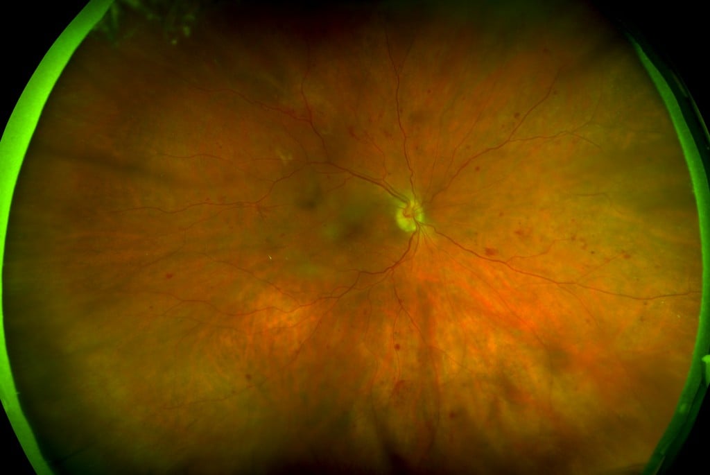
Image of proliferative diabetic retinopathy (DR) captured with the California
Many practitioners are rising to these challenges with the help of a technology that has unique, proven capabilities in improving DR diagnosis and management – ultra-widefield (UWF™) retinal imaging.
Ultra-widefield Retinal Imaging
called optomap. The optomap is produced in a single capture and does not require pupil dilation, but can be utilized as a complementary procedure. Along with UWF color imaging, UWF retinal imaging technology can also support UWF fluorescein angiography (FA), UWF fundus autofluorescence (FAF), and UWF indocyanine green chorioangiography (ICG).
UWF retinal imaging technology was pioneered and continues to be advanced by Optos. There are more than 8,700 Optos UWF retinal imaging systems in operation in the United States, the European Union, and elsewhere in the world. To date, these retinal imaging systems have produced more than 49 million optomaps.
Visualization of the Retinal Periphery for Improved Diagnostic and Treatment Outcomes
Multiple studies have confirmed that the diagnostic accuracy of UWF color imaging is comparable to conventional 7 standard field (7SF) ETDRS photographs. However, in several key areas, UWF improves the practitioner’s ability to identify and track the progression of DR.
— The ability to visualize the periphery of the retina can provide the practitioner with key insights into DR pathology and the potential for disease progression. One recent study examined patients using both 7SF and UWF. Over a four-year period patients with DR lesions outside of the normal 7SF field of view were found to be at four times the risk for progression to proliferative DR than patients without predominantly peripheral DR lesions.3
— UWF imaging enables the practitioner to make a more comprehensive assessment of patient DR pathology. Studies using UWF fluorescein angiography (FA) have reported that UWF-FA captures more pathology than conventional 7SF, including nonperfusion and neovascularization. In some cases, retinal pathology was uncovered that was not evident in 7SF images.4 Another study compared DR severity grading using UWF and 7SF, and reported that 15% of patients were graded with more severe DR with UWF retinal imaging.5
UWF imaging is also enhancing diagnosis and treatment in related diseases such as diabetic macular edema, and in other conditions including age-related macular degeneration, branch retinal vein occlusion, Stargardt’s disease, retinitis pigmentosa, and sickle cell retinopathy.
Another setting where digital imaging is improving diagnostic screening results is ocular telehealth. Recent research has demonstrated that inclusion of UWF retinal imaging in a screening program not only identified more pathology but also significantly reduced evaluation time and the proportion of ungradable images.
The Next Decade
The growing population of people afflicted with DR will challenge both optometrists and ophthalmologists alike.
— Optometric practices will serve more patients diagnosed with diabetes as well as more patients with undiagnosed diabetes. Routine, year-to-year DR screening and accurate diagnostic review will take on added importance.
— Comprehensive ophthalmic practices and retinal specialists will manage a growing DR caseload. Better diagnostic tools will be needed to understand and predict DR progression and to assess treatment outcomes, particularly as new therapeutic approaches and novel treatments improve the management of DR early in the disease process.
UWF retinal imaging will enable faster, more definitive DR screening, more accurate and comprehensive diagnoses, and greater opportunities for favorable patient outcomes.

[1] IDF Diabetes Atlas: Seventh Edition; Centers for Disease Control, 2015 Diabetes Statistics
[2] IDF Diabetes Atlas: Seventh Edition
[3] Silva PS, Cavallerano JD, Haddad NM, et al. Peripheral lesions identified on ultrawide field imaging predict increased risk of diabetic retinopathy progression over 4 years [published online ahead of print February 19, 2015]. Ophthalmology. doi:10.1016/j.ophtha.2015.01.008.
[4] Wessel MM, Aaker GD, Parlitsis G. et al. Ultra-wide-field angiography improves the detection and classification of diabetic retinopathy. Retina. 2012;32:785-791.
[5] Silva PS, Cavallerano JD, Sun JK, et al. Peripheral lesions identified by mydriatic ultrawide field imaging: distribution and potential impact on diabetic retinopathy severity. Ophthalmology. 2013;120(12):2587-2595.