April is Women’s Eye Health and Safety Month, which may, at first glance, seem a bit of a niche concern. However, according to the organization Prevent Blindness, women make up the majority of the 4.4 million Americans, age 40 and older, who are visually impaired or blind. More women than men have age-related macular degeneration, cataracts, and glaucoma. These numbers will only continue to increase in the years to come.
Although there are no cures for some of these diseases, many of the effects may be lessened through early detection and treatment. A 2015 survey found that one in four women had not received an eye exam in the past two years. Since women may not be aware that they are at greater risk than men of developing eye disease that could lead to serious vision impairment, practitioners can take the opportunity during Women’s Eye Health and Safety Month to stress the importance of paying attention to eye health and the importance of an annual eye exam.
A comprehensive eye exam should include a thorough examination of the retina, including an optomap, which is complementary to a DFE and an excellent tool for screening and for patient education. Because an optomap image can be obtained in less than ½ second, it leaves ample time for the practitioner to educate on eye health.
Dr. Germain Burke and Britta
Dr. Germain Burke, of Burke Optometry in Lodi, California, understands the value of optomap in communicating with and educating her patients. She recounts a story about her patient, Britta, that demonstrates how an optomap screening can reveal issues in asymptomatic patients, as well as, patients who have trouble communicating or who may minimize health issues or concerns.
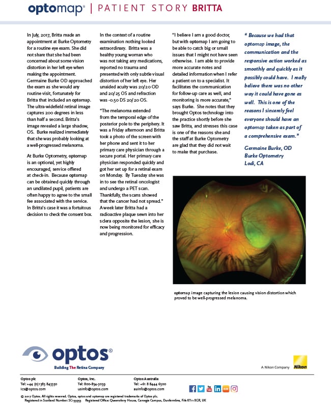
In July 2017, Britta made an appointment with Dr. Burke for a routine eye exam. She did not share that she had been concerned about some vision distortion in her left eye. However, Burke approached the exam as she would any routine visit, and fortunately for Britta, that included an optomap. The ultra-widefield digital retinal image provides a 200° view of the retina, out to the far periphery, and this .04 second capture revealed a large shadow OS. Burke realized immediately that she was looking at a well-progressed melanoma.
“The melanoma was in the periphery but very clear. It was a Friday afternoon so she took a picture of the optomap image on the screen and sent that immediately to her primary care physician through a secure portal. Her PCP responded quickly and got her set up for a retinal exam on Monday. On Tuesday she was in to see the oncologist and a PET scan, thankfully, showed that the cancer had not spread.” A week later Britta had a radioactive plaque sewn into the sclera opposite the lesion and is now being monitored for efficacy and progression.
“I believe I am a good doctor but with optomap I feel like I am going to be able to catch big or small issues that I might not have seen otherwise. I can provide more accurate notes and detailed information when I refer a patient on to a specialist. It facilitates the communication for follow-up care as well, and monitoring is more accurate,” says Burke.
She notes that they brought optomap technology into the practice shortly before she saw Britta, and stresses that this case is one of the reasons that she and the staff at Burke Optometry are glad that they did not wait to make that purchase. “Because we had that optomap image, the communication and the responsive action worked as smoothly and quickly as it possibly could have. I really believe there was no other way it could have gone as well. This is one of the reasons I sincerely feel everyone should have an optomap taken as part of a comprehensive exam.”
Visit our website to find a doctor such as Dr. Burke, who has optomap in their practice.