When 29-year-old Emmy came to see Uwe Canting, OD at Canting Optometry in Cary, NC, she was relatively certain that her eye was fine, but wanted to seek reassurance from her optometrist. Emmy had received a high-impact, full-blown soccer ball to the eye during a soccer match the preceding day and while having no symptoms other than slight discomfort from the bruising, she realized that the impact was severe enough that something unseen may have occurred.
Canting notes that Emmy presented with a black eye OD, while her visual acuity was 20/20. “The eye itself looked fine. Other than the ecchymosis, there were no immediate concerns. There was no apparent subconjunctival hemorrhage and no recession of the iris. But, while dilated, I could see instantly that it was not normal and decided to capture an optomap image. Sure enough, the image clearly showed the whitish sheen of Commotio retinae superiorly temporal.” Canting recalls, “The beauty of this situation was that I had her optomap image from her last visit and I could show her, clearly and tangibly, what had occurred in her eye.”
He adds that optomap has proved to be extremely valuable for patient education in a variety of scenarios because his patients love to be able to see what he sees and better understand their ocular health. “There is a wonderful opportunity to educate my patients when I use optomap to help explain the anatomy of their eye. If the patient is anxious, we may review the image right away. Otherwise I save it for the end of the exam. If there is an issue I can easily show them, better than it could be explained. Otherwise, it simply puts their mind at ease.” In Emmy’s case Canting could show her the pathology and stress why she should have further examination. Canting referred her immediately to the retinal specialist who reported later that he would be following Emmy’s condition to ensure that no subsequent issues developed.
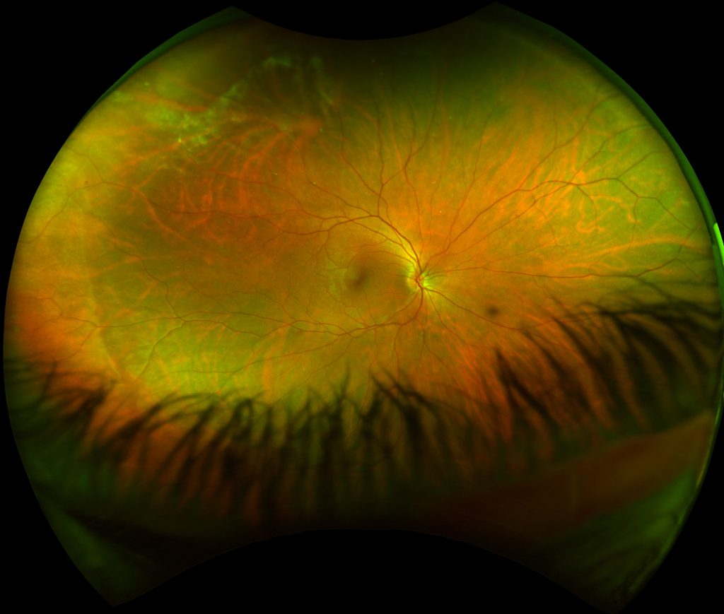
optomap image of Emmy’s right eye, post trauma showing evidence of Commotio retinae
Commotio retinae is a term that describes the damage that occurs to the outer retinal layers, caused by shock waves that surge through the eye from the impact site of a blunt trauma, such as Emmy’s soccer ball incident. Often the damage is recognized in the posterior pole but can also manifest, as it did for Emmy, in the periphery where it may be overlooked on a standard central pole retinal view. While most cases of the condition resolve spontaneously within a month to six weeks, more severe cases can cause temporary or permanent vision loss. Secondary issues can include choroidal neovascularization, retinal tears, detachment, zonular dehiscence, angle closure glaucoma and lens dislocation.
Rather than shrug off the soccer ball impact injury, Emmy was wise to seek a medical opinion and was fortunate that the injury was not more severe. Canting describes how optomap was a valuable tool in the discovery of the damage and the critical follow-up care that would give Emmy peace of mind. “She called me later, to tell me how grateful she was that I had seen the issue and had referred her to the specialist for further evaluation.”
As May is Healthy Vision Month, let’s all use Emmy’s story as a reminder to the importance of the often-overlooked recommendation encouraging protective eyewear for all ages in sports that could involve impact to the eye.
AAO recommends the following Daily Practices for eye health:
- Regular eye exams to detect subtle changes in the eye
- Wearing UV protection sunglasses
- Wearing safety glasses while playing impact sports or doing work about the yard/house that may present hazards to the eye
- Proper contact lens care
- Eating a healthy and balanced diet
- Cessation of smoking
Protect your vision – and visit an eyecare professional who uses optomap ultra-widefield retinal imaging in their practice!