Glaucoma is a degenerative, sight-threatening disease regarded as one of the major causes of blindness, accounting for an estimated 60 million people worldwide. By the year 2020 this number is thought to increase to around 80 million people globally.1
In a recent study2, the potential use of UWF imaging to detect glaucoma, and specifically to evaluate the reproducibility of measures of vertical cup-to-disc ratio (VCDR) using UWF, and the agreement between UWF and standard color digital stereoscopy (CDS), was conducted.
The purpose of this study was to evaluate the reproducibility and validity of UWF imaging in estimating VCDR measurements.
- Observational study
- 100 eyes from 100 consecutive patients using CDS and UWF
- Northern Ireland Cohort for the Longitudinal Study of Aging (NICOLA)
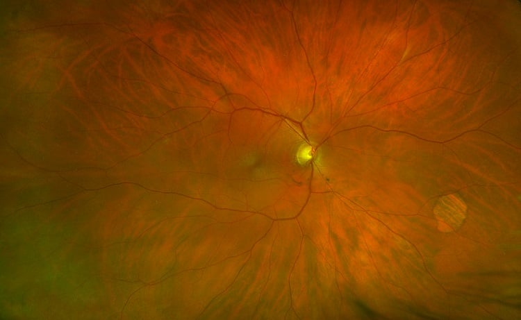
Glaucoma patient with optomap image (image courtesy of William Lesko, MD)
A factor to consider when estimating VCDRs using different ophthalmic techniques is the dimension of the image it produces. 3D v 2D, as well as image color affecting appearance for interpretation of cup depression and elevation and vessel contours. However, previous studies have reported on the value of non-stereo fundus images to evaluate disc cupping reporting no differences in diagnostic performance between monoscopic and stereoscopic images when detecting glaucoma.3,4
All color fundus disc photographs and UWF retinal images were graded by two masked trained observers and one masked glaucoma specialist. There was approximately one month between grading the first (CDS) and second (UWF) measurements to reduce the possibility of remembering previous data. Observers 1 and 2 regraded 50 optomap images to assess intra-observer reliability independently of each other. The outputs of both the cup and disc measurements were given in pixels. This was then divided in order to give the VCDR value.
The results of the study demonstrated almost perfect agreement between color digital stereoscopy and the optomap, an ultra-widefield imaging technique when assessed by a glaucoma specialist. It also showed the UWF technique was reproducible in VCDR estimates. Our data suggest that UWF imaging may be suitable for diagnosing glaucoma in situations in which slit-lamp biomicroscopy or digital color stereoscopy are not available. While clinical examination with slit-lamp biomicroscopy is the accepted gold standard, many clinics use digital methods for screening efficiency. Further research about the comparative diagnostic performance of UWF and other imaging technologies may be warranted. Read the full paper here
UItra-widefield optomap technology has been used in over 400 clinical research papers and peer-reviewed studies. Previous validation studies have also found clinical equivalence between UWF optomap imaging and traditional fundus imaging for the evaluation of diabetic retinopathy and age-related macular degeneration5,6. Find out more about the clinical outcomes from optomap technology, on our website.
- Quigley HA, Broman AT. The number of people with glaucoma worldwide in 2010 and 2020. Br J Ophthalmol. 2006;90(3):262– 10.1136/bjo.2005.081224.
- Can ultra-wide field retinal imaging replace colour digital stereoscopy for glaucoma detection? Quinn NB, Azuara-Blanco A, Graham K, Hogg RE, Young IS, Kee F.
- Hitchings RA, Genio C, Anderton S, Clark P. An optic disc grid: its evaluation in reproducibility studies on the cup/disc ratio. Br J Ophthalmol. 1983;67(6):356– 361. 10.1136/bjo.67.6.356
- Sharma NK, Hitchings RA. A comparison of monocular and ‘stereoscopic’ photographs of the optic disc in the identification of glaucomatous visual field defects. Br Ophthalmol. 1983;67(10):677–680. 10.1136/bjo.67.10.677.
- Silva et al. Nonmydriatic Ultrawide Field Retinal Imaging Compared with Dilated Standard 7-Field 35-mm Photography and Retinal Specialist Examination for Evaluation of Diabetic Retinopathy. American Journal Of Ophthalmology, 2012
- Agreement between image grading of conventional (45°) and ultra wide-angle (200°) digital images in the macula in the Reykjavik eye study. Eye (Lond). 2010 Oct;24(10):1568-75.