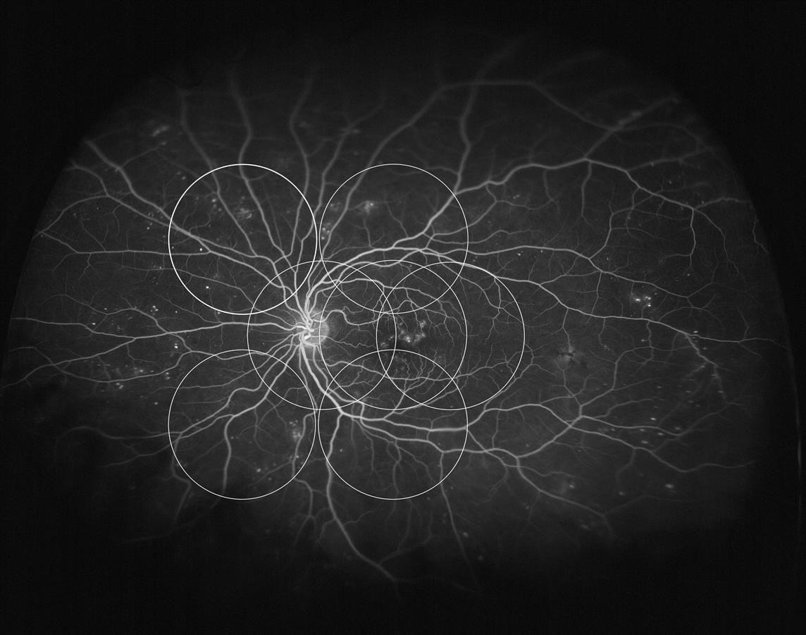
A study published in Diabetes Care comparing Optos nonmydriatic UWF imaging to mydriatic ETDRS 7-field photography in screening for DR found that the two methods showed considerable agreement in identifying clinically significant macular edema and grading DR level. However, UWF permitted visualization of a substantially larger retinal area, which may be advantageous in the diagnosis and management of DR. Because UWF does not require dilation, it also may be more acceptable to patients, thereby improving compliance with screening programs, and could facilitate remote image generation and interpretation through telemedicine.

optomap® fa demonstrating the peripheral changes associated with diabetic retinopathy that are not able to be visualized using ETDRS 7 standard field photography.
Kernt M, Hadi I, Pinter F, Seidensticker F, Hirneiss C, Haritoglou C, Kampik A, Ulbig MW, Neubauer AS. Assessment of diabetic retinopathy using nonmydriatic ultra-widefield scanning laser ophthalmoscopy (optomap) compared with ETDRS 7-field stereo photography. Diabetes Care. 2012: 1-5.