optomap® Recognizing Pathology
This material is designed as a searchable reference resource to support clinical decision-making. The information contained here should be used as general guidance when viewing optomap and OCT images from Optos devices. The differential diagnosis should be made under the direction of the responsible physician. These images were taken on the latest ultra-widefield optomap devices.
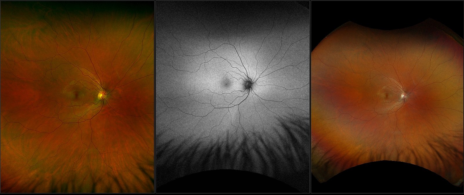
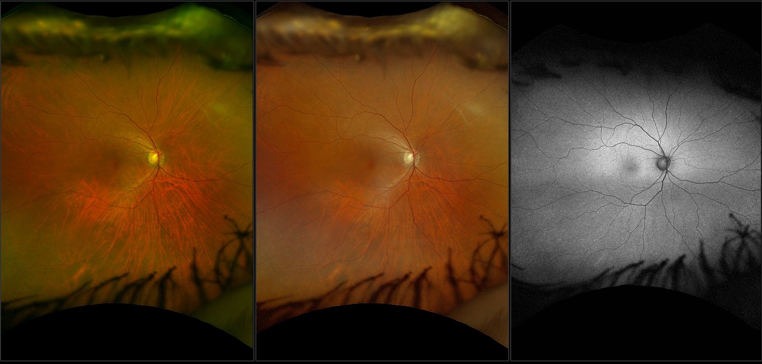
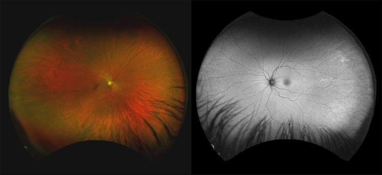
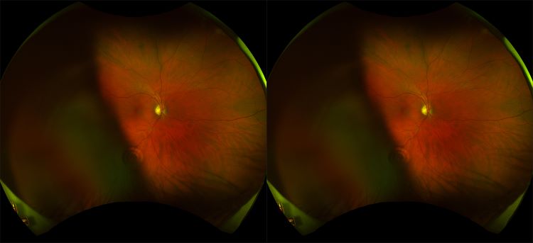
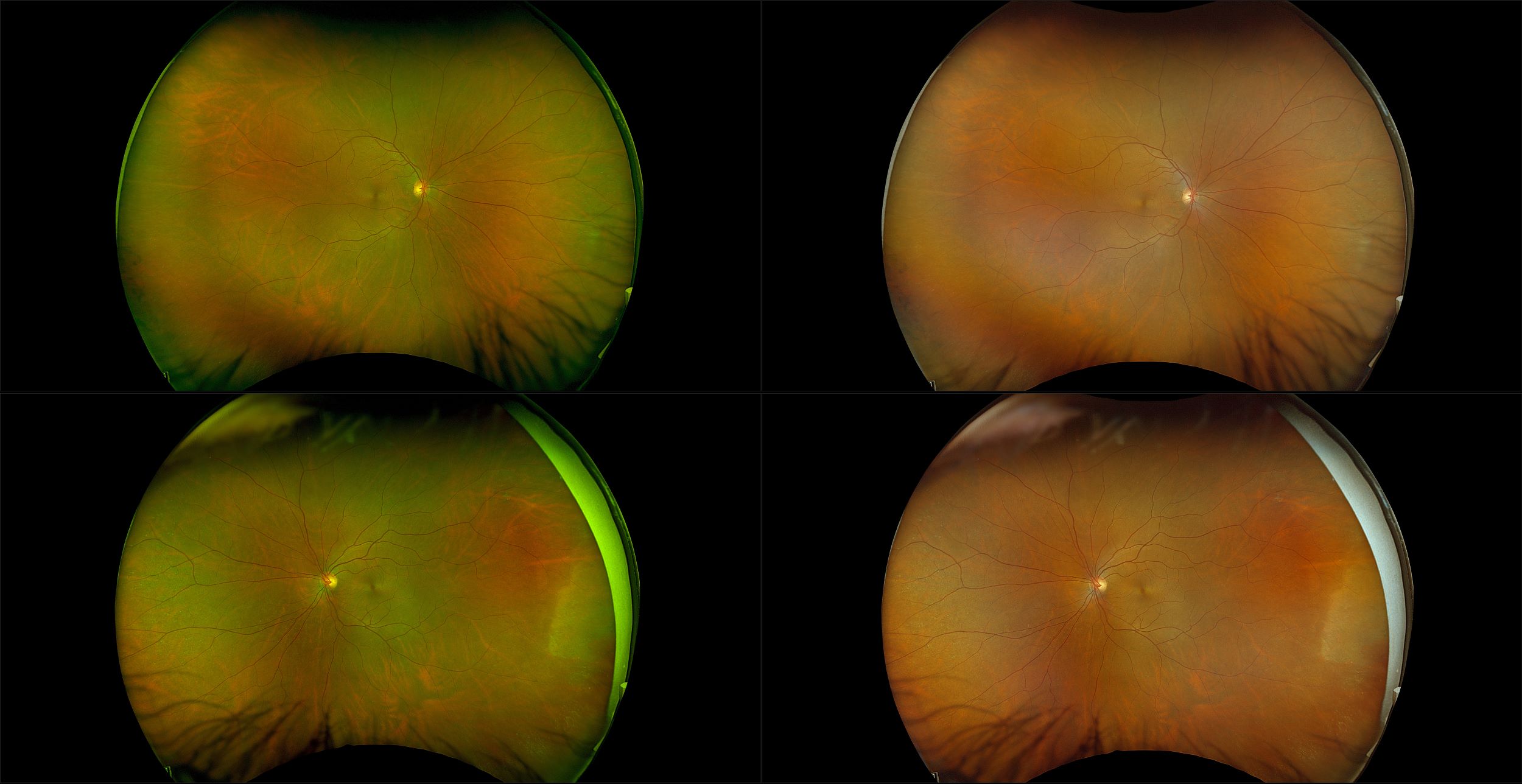
Healthy Retina
The retina is composed of the inner neural or sensory layers and the outer pigment epithelium. The neural retina has nine layers; from inner to outer they are the internal limiting membrane, nerve fiber layer, ganglion cell layer, inner plexiform layer, inner nuclear layer, outer plexiform layer, outer nu-clear layer, external limiting membrane, and photoreceptors. The normal retina is essential transparent (except for the pigment in the blood), although it does absorb a quantity of light passing through it. The sensory retina in the periphery is thin and relatively weak and thus is susceptible to full thickness breaks.