You are now leaving the Global site.

Ok Cancel
optos
  • Products
  • Patients
  • Providers
  • About Us
  • Contact
  • Customers
  • Language Menu
    English Deutsch Français
Home > Recognizing Pathology >

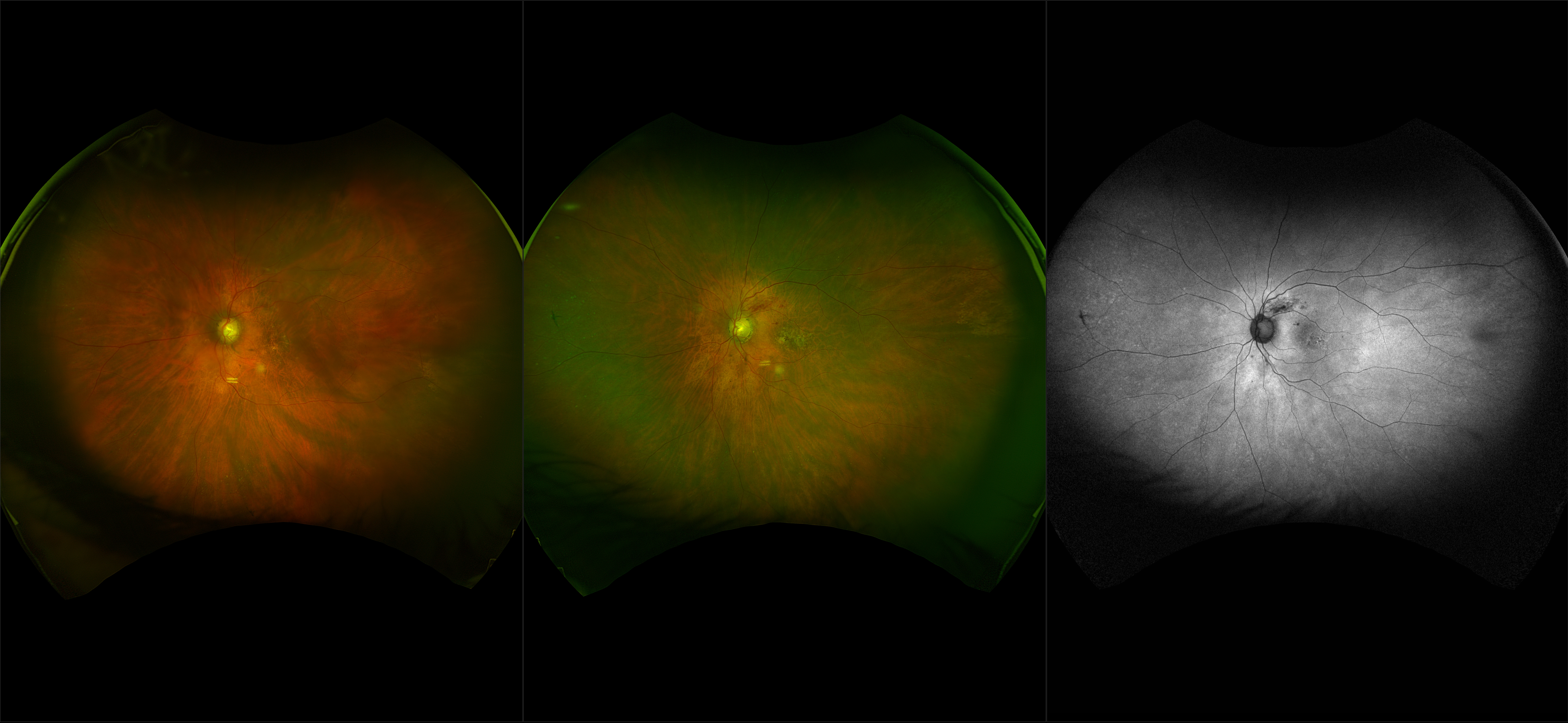
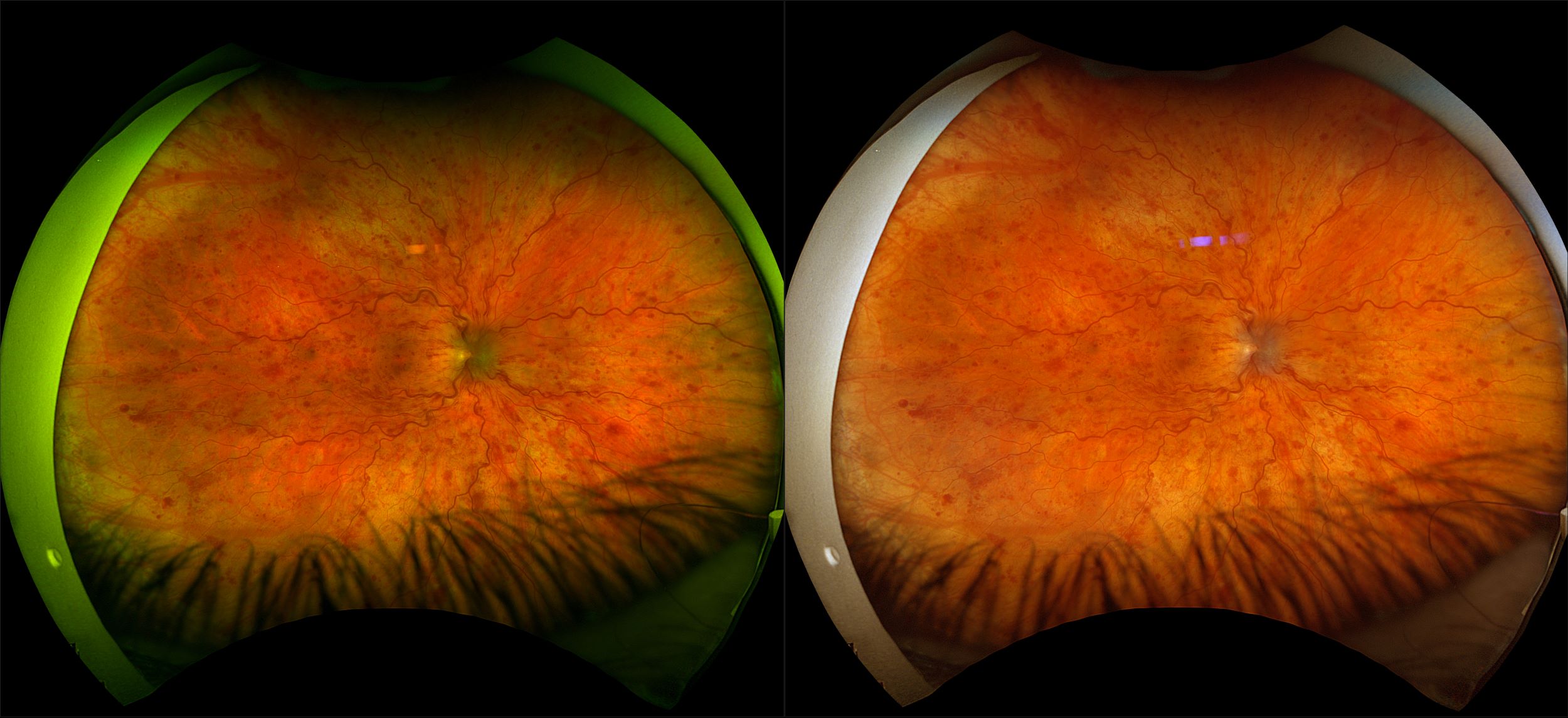
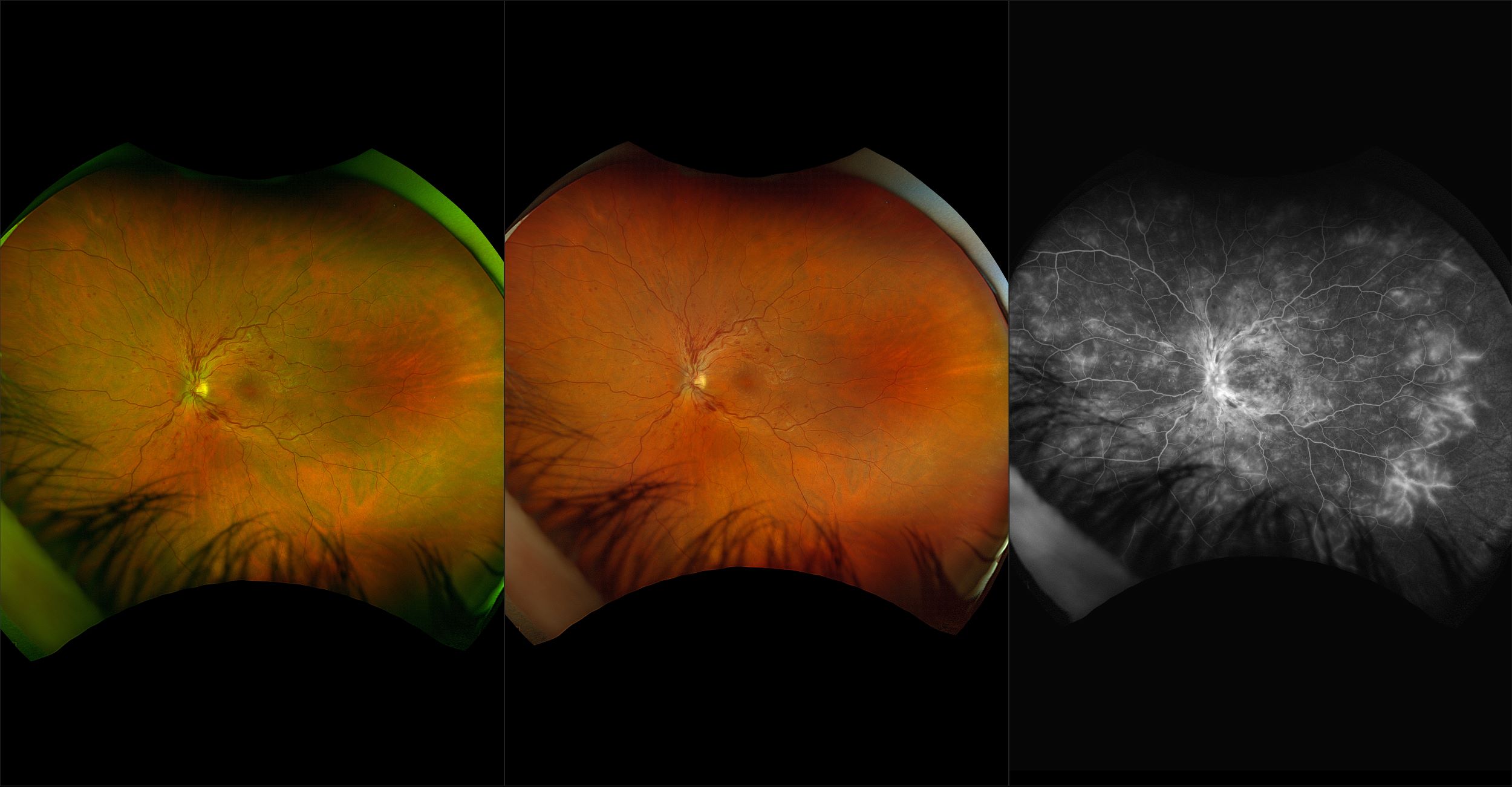
Central Retinal Vein Occlusion (CRVO)

Thrombosis of the central retinal artery or compression of the adjacent central retinal artery may cause a CRVO. Hemorrhages, venous dilation, and in some cases cotton-wool spots can be seen both in the posterior pole and periphery. There is retinal edema and sometimes optic disc edema. Sometimes there may be neovascularization of the retina or iris. This may require photocoagulation or anti-VEGF injection to attempt to achieve regres-sion of the new vessels. Neovascularization of the iris (NVI, rubeosis iridis) may lead to an anterior chamber bleed or neovascular glaucoma (this often occurs about 90 days later). When the drainage of the central retinal vein is slowed significantly, then preexisting vessels at the margin of the optic disc may shunt blood flow to the choroid (optociliary vessels). These shunt vessels appear as tortuous vessels red worms on or next to the disc.
View Full Case

Device

Image Modality

  • optomap
  • OCT

How can we help?

If you have questions regarding the cases, images, or pathologies, or if you would like further information about our ultra-widefield retinal imaging technology, please complete the form below.
I want to receive email updates about Optos products and services
<p>You are now leaving the Global site.</p> Ok Cancel
  • Blog
  • Cookie Policy
  • Careers
  • Distributor Portal
  • Manage Preferences
  • Media
  • News
  • Privacy
  • Registered in Scotland
  • UK Modern Slavery Act

© 2023 Optos. All rights reserved. Company Registration Number SC139953