Recent research suggests that patients suffering from various systemic disorders may have their disease state impacted by the addition of ultra-widefield (UWF™) retinal imaging to their examination. New research has found that patients with sickle cell disease may benefit from UWF retinal imaging for the diagnosis and management of sickle cell retinopathy (SCR).
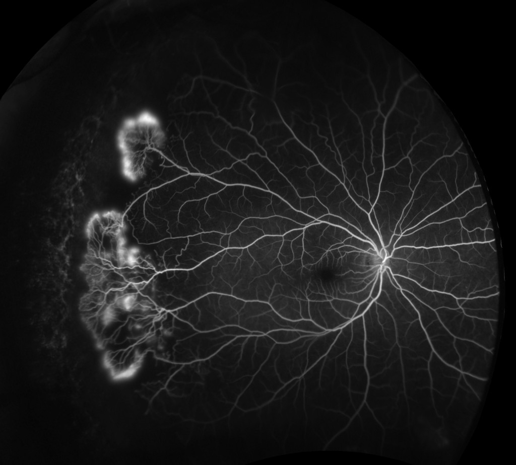
Sickle Cell Retinopathy FA
Background
Sickle cell disease is an inherited disorder in which the body produces blood cells with abnormally formed hemoglobin. Symptoms include anemia, severe and chronic pain, infection, hypertension, hand and foot swelling, leg ulcers, and retinal vascular changes1. Sickle cell disease can also have an impact on vision. Sickle cell retinopathy mainly affects the peripheral retinal vasculature2 as the result of abnormal, sickle-shaped blood cells becoming trapped in the small blood vessels of the eye3. Non-proliferative SCR, characterized by retinal hemorrhage from superficial blood vessels, can cause loss of visual acuity. Proliferative SCR is marked by vascular occlusions that lead to localized ischemia, neovascularization, and in later stages blindness from vitreous hemorrhage or tractional retinal detachment. Patients with sickle cell disease are at varying degrees of risk of developing SCR, but those with the type SC or S-Thal hemoglobin genotypes are at significant risk for developing proliferative SCR4. Traditionally ETDRS 7 standard field imaging has been used to try to manage and treat the disorder.
Ultra-widefield imaging technology is proving of unique value in visualizing SCR pathologies of the peripheral retina. UWF imaging technology enables the capture of a 200-degree view of the retina and gives ocular health practitioners imagery and diagnostic information that can’t be provided by conventional imaging methods. Starting with color (red and green) optomap imaging, Optos has systematically extended its UWF-based technology into a multi-modal platform that supports fundus autofluorescence (optomap af), fluorescein angiography (optomap fa) and indocyanine green angiography (optomap icg).
Screening for Sickle Cell Retinopathy Using UWF Imaging
A growing body of clinical data indicates that sickle cell disease compared the accuracy of UWF imaging with traditional clinical fundus examination. The researchers’ conclusions show that optomap color imaging is as sensitive as the clinical fundus exam in detecting SCR in children and adults; optomap images had revealed higher grades of SCR in many eyes as compared to the severity indicated by a clinical fundus exam; and optomap images proved more effective than clinical fundus exams in detecting early, non-proliferative SCR in children and adults.
UWF Imaging Correlation with Other Imaging Technologies
Fluorescein angiography is often used in SCR diagnosis and management, and a recent study6 evaluated the value of optomap fa (fluorescein angiography) by correlating optomap fa results with other imaging modalities.
This retrospective case study evaluated ten patients (18 eyes), with sickle cell disease who had undergone simultaneous optomap fa along with spectral domain optical coherence tomography (SDOCT) and optical coherence tomography angiography (OCTA). The authors observed that:
— optomap fa images identified varying levels of SCR in all 18 eyes.
— Higher levels of retinal ischemia (as estimated by a peripheral ischemic index calculated from optomap fa images), were well correlated with more severe levels of SCR.
— SDOCT found no remarkable pathology in seven eyes (38.9%) but, in other eyes, areas of macular atrophy were found that were correlated to a higher ischemic index.
The study concluded that a multimodal imaging approach, including UWF, could help more accurately characterize the progression of SCR.
Sickle Cell Retinopathy Management Using optomap fa
Researchers7 used optomap fa (fluorescein angiography), as well as conventional imaging techniques to diagnose and help guide the treatment of SCR in a group of six patients with sickle cell disease. Five of the six had type SC hemoglobin. The study patients reported no prior ocular complaints and, with the exception of one eye, all eyes had a central visual acuity of 20/20. Among the study’s findings:
— Fifty percent of the eyes (four patients), were found to have proliferative changes including one with Goldberg Stage 4 vitreous hemorrhage. The illustrates the need for close surveillance of at-risk patients regardless of symptomatic presentation.
— In three eyes (25%), conventional imaging missed peripheral vascular changes which were visible on UWF imaging. This led to changes in treatment for two of the eyes.
The researchers also observed that use of optomap fa improved patient management via easier detection of proliferative changes over a larger area of the retina and more precise calculations of the extent of retinal ischemia, which is a key risk factor in SCR progression.
* * *
Multimodal UWF imaging is providing practitioners with (1) a powerful screening tool for sickle cell retinopathy and, (2) a comprehensive diagnostic imaging method that can characterize the extent of SCR pathology and help assess the risk of progression. This promises to improve the detection and management of sickle cell retinopathy. UWF imaging is proving useful in the detection, management and treatment of many systemic diseases that have retinal manifestations.

Sources:
- https://emedicine.medscape.com/article/205926-overview
- Cho M, Kiss S., Detection and monitoring of sickle cell retinopathy using ultra wide-field color photography and fluorescein angiography. Retina. 2011 Apr;31(4):738-47. doi: 10.1097/IAE.0b013e3181f049ec.
- American Academy of Ophthalmology, Sickle Cell Retinopathy, https://eyewiki.aao.org/Sickle_Cell_Retinopathy
- American Academy of Ophthalmology, Sickle Cell Retinopathy, https://eyewiki.aao.org/Sickle_Cell_Retinopathy
- Lam, Alabduljalil, VandenHoven, et al, The use of retinal wide-field imaging system to screen for sickle cell retinopathy (SCR), Poster, ARVO 2016 Annual Meeting
- Ghasemi F., Scott AW, Wang K, et al, Correlation of Multimodal Imaging in Sickle Cell Retinopathy, Retina. 2016 Aug 4 [Epub ahead of print]
- Cho M, Kiss S., Detection and Monitoring of Sickle cell Retinopathy using Ultra Widefield Color Photography and Fluorescein Angiography, Retina. 2011 Apr;31(4):738-47. doi: 10.1097/IAE.0b013e3181f049ec.