When Vince Young, OD introduced the Daytona from Optos in his practice, he volunteered to be the imaging guinea pig while his staff was being trained on the device. He was unnerved when he reviewed his images with the trainer and somewhat uncertain about what he was actually seeing. He knew what a posterior subcapsular cataract (PSC) looked like through the slit lamp but was surprised by what the optomap image laid evident. While optomap is known for being able to penetrate through medial opacities far better than white light modalities, a PSC, which tends to be denser than other types of cataract, will cast a shadow on the retina revealing the issue. A concerned Dr. Young sent the image to his wife, Lindsey Brewer Young, OD. When she reviewed the image on her phone she immediately responded, questioning whose eye she was regarding. Learning it was her husband’s image she returned to the clinic, conducted a dilated exam, and confirmed that it was indeed a PSC that had been revealed in the optomap image.
Read the entire story by clicking on the image
Young, who is 40, had no reason to suspect he would have cataracts which are more typically associated with the elderly population. A subcapsular cataract occurs at the back of the lens and can be caused by a variety of circumstances such as systemic issues and some forms of medication. Posterior subscapsular cataracts are also more difficult to remove, due to adhesion of the cataract to the lens capsule, and there is an increased risk of capsule rupture during removal.1 However, Young’s cataract surgery on both eyes was successful and his vision is fine.
This discovery has become an excellent tool in communicating the importance of optomap to his patients. The image hangs in the exam room while Young and his staff frequently share the story. “If the doctor didn’t know he had a cataract, how would anyone else know?” they say, underscoring how valuable the screening can be. This unusual story has assisted with the elevated level of acceptance the optomap receives in the office.
Blanchard Eye Care sits in a quiet community outside Blanchard, OK and Young was concerned that he would have difficulty getting people in the rural community to embrace the technology. He, however, was convinced that if he at least broke even it would be worth the investment based on the clinical value. “Before purchasing the Daytona, I asked several of my colleagues about their experiences and they referred to it as a ‘no—brainer’,” says Young. He stresses that while the technology has without a doubt brought a financial bonus, the value of what it provides clinically far outweighs the monetary gain.
Young stresses how the exam experience has changed with optomap in an extremely valuable way when it comes to patient education. The optomap image enables him to help his patients understand exactly what is occurring in their eye, or even just to provide reassurance that all is well. “Even if there is no pathology, patients want me to take the images every year. They want to see what I see.”
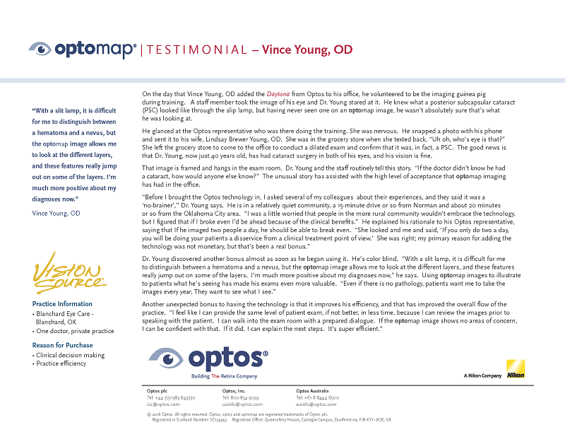
Young happens to be color blind and further identifies another contribution that optomap brings to him, personally. Because of this, a slit lamp view can sometimes make it difficult for him to distinguish between a hematoma and a nevus. “However, the optomap image allows me to look at the different channels, and these features really jump out on some of the layers. I’m much more positive about my diagnoses now.”
Dr. Young’s story demonstrates that it is not only possible for practitioners to image themselves and discover retinal pathology but also discover if significant opacities reside in the media as well.
Today, cataracts affect more than 22 million Americans age 40 and older. And as the US population ages, more than 30 million Americans are expected to have cataracts by the year 2020.2 optomap technology is being increasingly utilized in cataract surgery clinics for immediate views of the retina. The ultra-widefield view is obtained through problematic, medial opacities, where white light has difficulty, revealing any retinal issues that might be a concern prior to surgery, as well as, following surgery. The ability to quickly and easily observe and document retinal health before and after cataract surgery provides both the patient and practitioner a tremendous peace of mind.
2 https://www.allaboutvision.com/conditions/cataracts.htm