Ultra-widefield (UWF™) technology supports and enables practice efficiency for eyecare professionals across all settings1. The integration of optomap® technology as a routine diagnostic and screening tool has shown to improve workflow and increase service capacity within a very short time, according to many eyecare professionals. Its ease of use makes for a smooth implementation process, and clinically, optomap has been found valuable as documented in over 400 peer-reviewed papers. Further, optomap can facilitate timely referrals for clinical opinions, supporting earlier treatment interventions and promoting collaboration between a variety of healthcare professionals and eyecare professionals alike. These all equate to improved patient workflow, clinical accuracy, and timely diagnosis and treatment for patients2.

Practice Efficiency in all Eye Care Settings
In an attempt to improve patient flow in his practice, optometrist David Anderson (Miamisburg Vision Care, Ohio) invested in a Daytona from Optos and, found his expectations exceeded for efficiency and as a diagnostic tool. Dr. Anderson noticed that the addition of the optomap allowed for more proficient patient service, from start to finish. Likewise, ophthalmologist, Scott Segal, MD started using optomap imaging in his practice (Pasadena Eye Associates, Texas) four years ago. Within the first two weeks, it became an integral part of his practice and had positively impacted how he practiced medicine.
For the ophthalmology practice of Paul E. Tornambe (Retina Consultants, California), first-year incorporation of UWF into the practice resulted in a 4.4% increase in patient volume over the previous, which translated to $40,000 in incremental revenue; the second year by 7%. Ultimately, the total incremental revenue realized with the integration of UWF equated to roughly $76,000 per year.
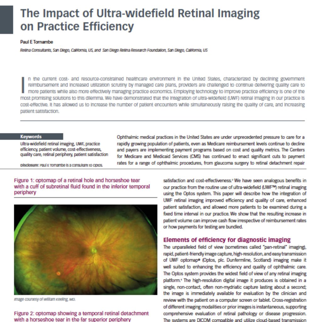
Per Dr. Segal, from a practice efficiency standpoint, optomap technology is able to capture a “wealth of information in the shortest amount of time3.” The UWF™ imaging capability of the optomap has “…proven exceptional for acquiring images of peripheral pathology.” In less than a second, optomap is able to generate a 200° (82%), accurate, high-resolution image of the retina, through an undilated pupil in a single capture. From a clinical outcomes vantage point, optomap offers multi-modal functionalities that are able to characterize subtle retinal pathologies, which may have been missed using traditional retinal imaging modalities.
optomap provides consistency for accurate documentation, disease tracking and pre- and post-treatment imaging, across all eye care settings. By using UWF imaging, it is possible to follow disease progression over time, and when patients require a treatment such as photocoagulation, it allows the doctor to target specific areas rather than using the less effective type of blanket laser coverage.
Many eyecare professionals comment on the ease of use of optomap. Operating this technology requires minimal training, and images can be captured quickly and efficiently and then networked to a consulting room, ready for review. Optometrist Aaron Werner has a practice in San Diego where he utilizes his Daytona as part of his “pre-testing protocol with all patients”. This means that their images are ready to view as soon as the patients walk into the examination room. He adds that “The Daytona allows us to do a quick and thorough screening and assessment of the retina and decide if it requires further attention.” optomap facilitates a streamlined approach, and according to Dr. Anderson, “…allows patients to be seen in one seamless swoop.”
Dr. Anderson and his team encourage all patients to have optomap imaging. His practice has a 92% opt-in rate, which has had a significant, positive impact on patient flow, boosting practice capacity an extra five patients per day. With this increase in patient flow, there has been a correlated rise in pathology detection, particularly in asymptomatic patients.
UWF images can be transmitted in a matter of seconds, which makes for prompt clinical decision-making and disease management. Ophthalmologist Dr. Nikolas London and Dr. Werner both practice in the San Diego, California, area, and both use UWF imaging. They understand the value of working together to benefit their patients. Dr. Werner is confident that, should he find anything that may warrant referral, he “…can send him [Dr. London] a picture of the retinal image and get quick feedback.” If Dr. London feels the condition is urgent, they can “…start formulating a treatment plan before the patient even walks through his door.”
As a practice efficient technology, optomap is a win-win option for all eyecare professionals and their patients and, according to Dr. Tornambe, it delivers “…clinically-relevant information, more rapidly, in a more patient-friendly manner, and can make important contributions to multiple aspects of the efficiency challenge.”
Increase patient throughput and practice efficiency in less than half a second, with optomap. Visit our website to learn more!
References
- Brian Kim MD, Kenneth A. Fukuda, OD, Karen P. Skvarna, OD – Ultra-Widefield Retinal Imaging Facilitates Integrated Eye Care. Use of optomap can identify more pathology and support efficient collaboration. Advanced Ocular Care, March 2017
- Paul E. Tornambe, The Impact of Ultra-Widefield Retinal Imaging on Practice Efficiency, USOR 2017;10(1):27–30
- Nikolas London, MD, Aaron Werner, OD, Tech Spotlight: Optos Ultra-Widefield Imaging Devices, July 6, 2016 – https://www.opthalmologyweb.com/Tech-Spotlights/188712-Optos-Ultra-Widefield-Imaging-Devices/