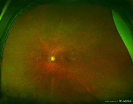
As we shared a few months ago, a study published in Diabetes Care comparing Optos’ nonmydriatic UWF imaging to mydriatic ETDRS 7-field photography as screening tools for DR showed UWF imaging as having an advantage in screening for DR. As Healio recently reported, UWF imaging is also considered “a viable option that may improve the efficiency of diagnostics for [DR] and diabetic macular edema.”
Jan Lammer, MD, shared at the Advanced Retinal Therapy Meeting that in addition to the standard methods of assessment and follow up for DR and diabetic macular edema (DME) – which include fluorescein angiography, fundus photography and others – Optos’ UWF imaging was utilized in a study of 206 eyes of 103 patients. Optos’ UWF imaging was used because it allowed practitioners to “detect peripheral alterations of the retina that would otherwise go undetected.”
The study compared the results of UWF imaging with conventional non-mydriatic fundus photography (NMFP). While the UWF images identified similar results in terms of rates and severity of DR as NMFP, it also “identified additional peripheral lesions” in over 24 percent of patients, which Lammers said may have indicated more severe cases of retinopathy severity grading in about 10 percent of the eyes studied.
Additionally, the study revealed that UWF imaging identified DR more frequently than NMFP. Practitioners also reported that their time spent evaluating images decreased by 28 percent with UWF images.
Are you interested in learning more about the role of UWF imaging in the assessment and treatment of DR? Read our blog post, “UWF imaging impacts assessments of severity in diabetic retinopathy (DR),” as well as case studies available on our website for more information.
If you’re interested in bringing our UWF imaging technology into your practice, request a consultation with a representative.
