Choroidal nevi are commonly reported as incidental findings in asymptomatic patients during routine eye exams. Nevi on the retina, similar to “freckles” on the skin, should be monitored for changes as they may turn in to melanoma1.
With the increased use of detection of choroidal neoplasms and nevi2.
Dr. Bryan Stoller reported using optomap to monitor Jeffry, a 54 year-old man with glaucoma3. Jeffry had his intra-ocular pressure (IOP) checked every four months, and returned annually for UWF imaging of his optic nerve and a small choroidal nevus. During one visit, while using optomap af, Dr. Stoller noticed that the nevus had started to autofluoresce. Although the choroidal nevus, measuring three disc diameters (DD), had not increased in size, the hyperfluorescence of the lipofuscin gave Dr. Stoller cause to investigate further. A dilated, cross-sectional OCT of the retina revealed that the nevus was significantly elevated, prompting Dr. Stoller to make an immediate specialist referral.
Dr. Stoller also noted that since using optomap technology, there had been a rise in the discovery of previously unobserved retinal pathologies in his practice. As a result, he increased his use of optomap af for all patients presenting with choroidal nevi. Dr. Stoller believes that optomap af facilitates earlier diagnosis, direct treatment and more successful patient outcomes.
A patient named George was referred to Dr. Albert Morier by his primary care physician to be monitored for diabetic progression. George’s ophthalmologist had also made him aware that he had a ‘freckle’ on his retina4. Dr. Morier dilates all patients who have a pre-existing medical condition, utilizing optomap as complementary to the dilation. Unfortunately, George did not dilate well and UWF imaging proved vital in his case. Along with trace diabetic retinopathy, Dr. Morier noted that the ‘freckle’ was actually a large lesion that had extended beyond the choroid into the retina. A referral was expedited for further assessment, and malignancy was confirmed.
In general, Dr. Morier has noticed that the number of asymptomatic patients, who have been imaged on the optomap, are increasingly presenting with ocular pathologies. He emphasized that, “It is knowing that likely I will not have missed something with the ultra-widefield view that optomap provides and that allows me to sleep better. It is really this that matters – quality of care – and peace of mind.”
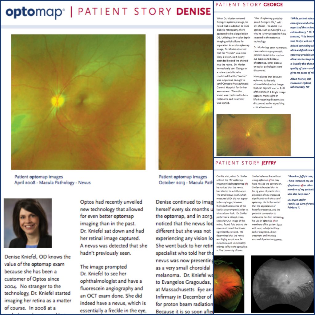
Denise Kniefel, OD has been using Optos technology since 2004. In 2008 her her optomap image showed the presence of a small nevus. She had been monitoring it herself on a regular basis, and in 2013, she noticed a slight change.
She immediately contacted a specialist who confirmed that the nevus had converted into a melanoma. She sought additional treatment at Massachusetts Eye and Ear and attributes the early detection of the change as key to her ongoing and successful treatment. Dr. Kniefel’s personal experience has compelled her to recommend UWF imaging to all her patients and those within her personal community5.
As highlighted above, and in other similar cases in general, the proactive, routine use of UWF imaging provides a complementary, comprehensive diagnostic modality to support early detection, timely referral and treatment of retinal pathology, particularly in asymptomatic patients. optomap technology supports improved patient outcomes, with greater patient participation, compliance and satisfaction. To find out more about optomap and to find an eyecare professional who uses optomap in their practice, please visit our website.
Sources:
1-2. Mark R. Kapperman, OD. Taking Retinal Imaging to the Next Level. Consider ultra-widefield imaging as a practice enhancer. Advanced Ocular Care. Cover Focus. November/December 2015. https://eyetubeod.com/2015/12/taking-retinal-imaging-to-the-next-level
3. Patient story – Jeffry: https://blog.optos.com/wp-content/uploads/2017/08/Patient-Story-Jeffry-1-1.pdf
4. Patient story – George: https://blog.optos.com/wp-content/uploads/2017/08/Patient-Story-George-1-1.pdf
5. Patient story – Denise: https://blog.optos.com/wp-content/uploads/2017/08/Patient-Story-Denise-Portrait-1-1.pdf