Jessica describes the events of that October 2017 day as somewhat serendipitous, even though what transpired illuminated a hidden threat to her life. Jessica, an actress, had just started rehearsing for a play in Billings, Montana, when she decided that contacts, rather than glasses, would better suit her part. “Really, I just thought it would be a good idea to be able to see while I was on stage,“ she laughs. “That’s all I needed was to get fitted for contacts. I didn’t feel that I needed, nor did I have time for, an eye exam. Besides I absolutely despise being dilated.” However, as fate would have it, she went that day to see Tom Felstet, OD, who feels strongly that a thorough view of the retina should be a part of every eye exam. Accordingly, Felstet had made it a priority to purchase an optomap ultra-widefield (UWF™) imaging device when he opened his new practice four years ago. He had the opportunity to utilize the technology during medical school and during his early years in practice. optomap is the only technology that captures over 80% of the retina in a single image, and it does so in a fraction of a second through an undilated pupil. When patients come to see Felstet he explains to them that it is important to have a comprehensive eye exam. He gives them the option to be dilated or, for a small fee, to have optomap screening without dilation. “Like many practitioners, I have always told my patients that they need to have a dilated exam at least every other year.” he explains. However, he stresses that changed after his experience with Jessica. “Now, it’s every year. I give all my patients the choice of dilation or an optomap exam. Over the past few years utilizing optomap I have realized that I discover far more pathology, often far sooner, than I might without it. I have found changes and pathology, that I could have missed otherwise.”
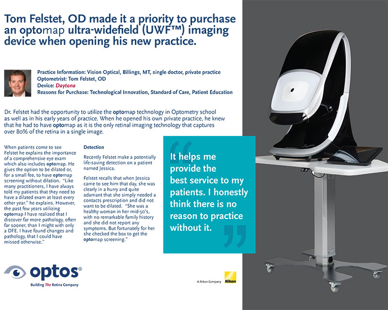
Felstet recalls that when Jessica came to see him that day, she was clearly in a hurry and quite adamant that she simply needed a contacts prescription and did not want to be dilated. “She was a healthy woman in her mid-50’s, with no remarkable family history and she did not report any symptoms. But fortunately for her she checked the box to get the optomap screening.”
When Dr. Felstet walked in to see Jessica he took one look at her image up on the screen and saw very clearly that there was a lesion in her right eye. “It was just far enough out that it would have been missed on an undilated slit lamp examination,” notes Felstet. When he showed it to Jessica he explained the area of concern and indicated that she should see an ophthalmologist. “He was very discreet,” Jessica recounts. “I know he did not want to alarm me. I could see quite clearly what he was talking about on the image, but even then, I was not really worried.” Jessica recalls that even when she did see the ophthalmologist, and he diagnosed the pathology as a choroidal melanoma, she still had difficulty accepting the gravity of the situation. “I mean, who had ever heard of a melanoma of the eye? That wasn’t even something on my radar. I had noticed some little flashes of light, but they were insignificant, and I just passed it off as reflections from some new glasses. Besides, in all other respects I was quite healthy.”
Jessica was then referred to an ocular oncologist in Denver and it was not until then that she grasped the significance of what was occurring. “My tumor was 11 mm wide, so it was right on the cusp – just ½ mm from what would have resulted in an automatic enucleation. Jessica was treated immediately and successfully with radioactive plaque therapy, but during surgery the tumor was biopsied revealing that she was genetically at high risk for metastases, particularly to the liver. Ocular melanoma tends to be aggressive and metastasizes, or spreads to other organs in the body, in about half of all cases.
Felstet says that he still gets chills when he thinks of how close Jessica came to losing her eye and how disruptive this experience was for him as a practitioner. He underscores that he has always been committed to providing comprehensive eye care, however he gives extra priority to communicating to his patients the critical importance of having a thorough examination of the retina every year.
“It is interesting to me how close that tumor was to her macula and yet she was not aware of any changes to her vision in that field, if she had not come in for her contacts when she did and if we had allowed her to refuse dilation, or an optomap, she would have likely lost her eye or could have lost her life.” He muses, “She returned to me recently and we took another optomap image, in looking at it I think that it is amazing that she is still here but that she also still has her central vision. Looking at that image again, I told her that she is miracle.”
Felstet notes that he has detected numerous pathologies since Jessica’s visit, from subtle pigment changes that suggested dangerous lesions, to BRVO in young, outwardly healthy individuals. “I am much more direct now about the importance of getting an optomap and about what I see on those images. I really would not want to practice without it because it would be hard to miss something as the pathology really stands out. It gives me enormous peace of mind.” Felstet is confident that optomap UWF will become the gold standard of care. “It helps me provide the best service to my patients. I honestly think there is no reason to practice without it.”
Read Dr. Felset’s full testimonial here…
Optos is committed to educating all on the importance of having regular eye exams. Protect your eye health by making optomap part of your yearly comprehensive eye exam. Visit our website for more sight-saving stories like Jessica’s and to find an optomap provider near you to schedule your eye exam today!