Introduction
Duke University Eye Center has been using their optomap enabled device since September 2006. During that time the image quality, increased field of view and ease of use have added value to the practice. Below details a case study which demonstrates the unique value of ultra-widefield fluorescein angiography offered by Optos in patients with age related macular degeneration.
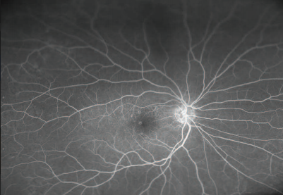
optomap fa image demonstrating both central and peripheral drusen consistent with AMD.
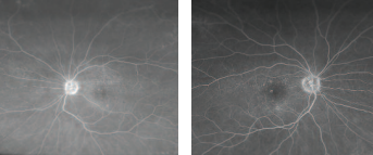
optomap fa images demonstrating excellent detail of the central macula in dry AMD (magnified images).
History
The female patient was diagnosed with dry AMD 1.5 years ago with nuclear sclerotic cataracts in both eyes. At this visit the patient presented with a change in visual acuity in the prior six months. Routine examination revealed an IOP of 15 in the right eye and 18 in the left, with visual acuity at 20/100(rt) and 20/40(lt).
Examination
This case study captured by the Optos ultra-widefield digital imagingdevice demonstrates the benefits offered by the technology. Duke University photographers were able to acquire a clear image set through relatively dense cataracts; something that is always a challenge with traditional mydriatic white-light based cameras. The red/green laser allows for less scatter as the beams enter the anterior segment allowing for a better image of the retinal surface.
The ultra-widefield image illustrates AMD changes affecting both the central and peripheral retina. Recent studies have demonstrated the peripheral retinal findings in early AMD, and this may be an important predictive phenotypic marker for outcomes.
The patient was reassured to know that she did not have wet AMD.
Conclusion
In just a short period, the Optos device has changed the way fluorescein angiograms are done within the practice. The capture of the 200° field in one image allows for a full diagnosis of retinal disease to be made easily because the progression of the dye across the retinal surface is caught at every stage. I believe ultra-widefield digital imaging will come to revolutionize the way eye care centers diagnose, triage and treat these cases.
For more information about this case studies and others, reach out to us today.
Author: Ivan J. Suñer, MD, Associate Professor – Duke University Eye Center